3G6N

Crystal structure of an EfPDF complex with Met-Ala-Ser


Experimental Data Snapshot

  • Method: X-RAY DIFFRACTION
  • Resolution: 2.50 Å
  • R-Value Free: 0.247 
  • R-Value Work: 0.225 
  • R-Value Observed: 0.225 

wwPDB Validation   3D Report Full Report


This is version 1.2 of the entry. See complete history


Literature

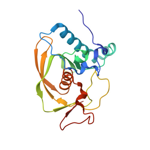
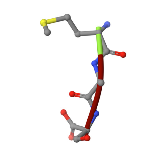
Crystal structure of an EfPDF complex with Met-Ala-Ser based on crystallographic packing.

Nam, K.H.Kim, K.H.Kim, E.E.Hwang, K.Y.

(2009) Biochem Biophys Res Commun 381: 630-633

  • DOI: https://doi.org/10.1016/j.bbrc.2009.02.113
  • Primary Citation of Related Structures:  
    3G6N

  • PubMed Abstract: 

    PDF (peptide deformylase) plays a critical role in the production of mature proteins by removing the N-formyl polypeptide of nascent proteins in the prokaryote cell system. This protein is essential for bacterial growth, making it an attractive target for the design of new antibiotics. Accordingly, PDF has been evaluated as a drug target; however, architectural mechanism studies of PDF have not yet fully elucidated its molecular function. We recently reported the crystal structure of PDF produced by Enterococcus faecium [K.H. Nam, J.I. Ham, A. Priyadarshi, E.E. Kim, N. Chung, K.Y. Hwang, "Insight into the antibacterial drug design and architectural mechanism of peptide recognition from the E. faecium peptide deformylase structure", Proteins 74 (2009) 261-265]. Here, we present the crystal structure of the EfPDF complex with MAS (Met-Ser-Ala), thereby not only delineating the architectural mechanism for the recognition of mimic-peptides by N-terminal cleaved expression peptide, but also suggesting possible targets for rational design of antibacterial drugs. In addition to their implications for drug design, these structural studies will facilitate elucidation of the architectural mechanism responsible for the peptide recognition of PDF.


  • Organizational Affiliation

    Korea University, Seoul, South Korea.


Macromolecules
Find similar proteins by:  (by identity cutoff)  |  3D Structure
Entity ID: 1
MoleculeChains Sequence LengthOrganismDetailsImage
Peptide deformylase
A, B
191Enterococcus faeciumMutation(s): 0 
EC: 3.5.1.88
UniProt
Find proteins for Q842S4 (Enterococcus faecium)
Explore Q842S4 
Go to UniProtKB:  Q842S4
Entity Groups  
Sequence Clusters30% Identity50% Identity70% Identity90% Identity95% Identity100% Identity
UniProt GroupQ842S4
Sequence Annotations
Expand
  • Reference Sequence

Find similar proteins by:  Sequence   |   3D Structure  

Entity ID: 2
MoleculeChains Sequence LengthOrganismDetailsImage
peptide (Met)(Ala)(Ser)C [auth F],
D [auth G]
3N/AMutation(s): 0 
Sequence Annotations
Expand
  • Reference Sequence
Experimental Data & Validation

Experimental Data

  • Method: X-RAY DIFFRACTION
  • Resolution: 2.50 Å
  • R-Value Free: 0.247 
  • R-Value Work: 0.225 
  • R-Value Observed: 0.225 
  • Space Group: P 64 2 2
Unit Cell:
Length ( Å )Angle ( ˚ )
a = 148.688α = 90
b = 148.688β = 90
c = 141.902γ = 120
Software Package:
Software NamePurpose
HKL-2000data collection
CNSrefinement
HKL-2000data reduction
HKL-2000data scaling
CNSphasing

Structure Validation

View Full Validation Report


Entry History 

Deposition Data

Revision History  (Full details and data files)

  • Version 1.0: 2009-03-03
    Type: Initial release
  • Version 1.1: 2011-07-13
    Changes: Version format compliance
  • Version 1.2: 2023-11-01
    Changes: Data collection, Database references, Derived calculations, Refinement description