3G6D

Crystal structure of the complex between CNTO607 Fab and IL-13


Experimental Data Snapshot

  • Method: X-RAY DIFFRACTION
  • Resolution: 3.20 Å
  • R-Value Free: 0.263 
  • R-Value Work: 0.213 
  • R-Value Observed: 0.213 

wwPDB Validation   3D Report Full Report


This is version 1.3 of the entry. See complete history


Literature

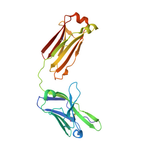
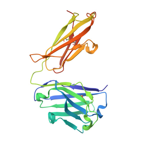
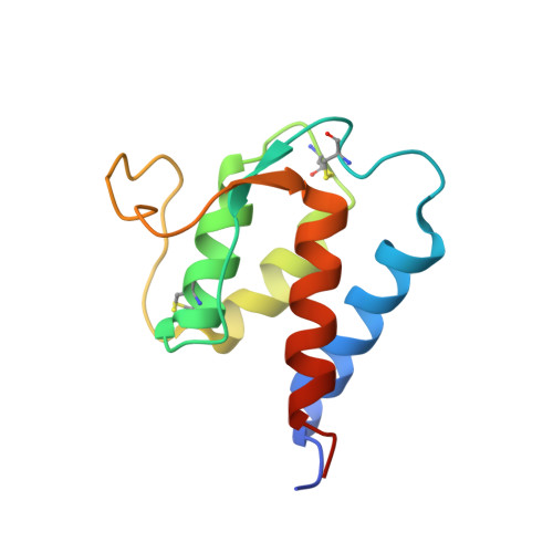
Epitope mapping of anti-interleukin-13 neutralizing antibody CNTO607.

Teplyakov, A.Obmolova, G.Wu, S.J.Luo, J.Kang, J.O'Neil, K.Gilliland, G.L.

(2009) J Mol Biol 389: 115-123

  • DOI: https://doi.org/10.1016/j.jmb.2009.03.076
  • Primary Citation of Related Structures:  
    3G6A, 3G6D

  • PubMed Abstract: 

    CNTO607 is a neutralizing anti-interleukin-13 (IL-13) human monoclonal antibody obtained from a phage display library. To determine how this antibody inhibits the biological effect of IL-13, we determined the binding epitope by X-ray crystallography. The crystal structure of the complex between CNTO607 Fab and IL-13 reveals the antibody epitope at the surface formed by helices A and D of IL-13. This epitope overlaps with the IL-4Ralpha/IL-13Ralpha1 receptor-binding site, which explains the neutralizing effect of CNTO607. The extensive antibody interface covers an area of 1000 A(2), which is consistent with the high binding affinity. The key features of the interface are the charge and shape complementarity of the molecules that include two hydrophobic pockets on IL-13 that accommodate Phe32 [complementarity-determining region (CDR) L2] and Trp100a (CDR H3) and a number of salt bridges between basic residues of IL-13 and acidic residues of the antibody. Comparison with the structure of the free Fab shows that the CDR residues do not change their conformation upon complex formation, with the exception of two residues in CDR H3, Trp100a and Asp100b, which change rotamer conformations. To evaluate the relative contribution of the epitope residues to CNTO607 binding, we performed alanine-scanning mutagenesis of the A-D region of IL-13. This study confirmed the primary role of electrostatic interactions for antigen recognition.


  • Organizational Affiliation

    Centocor R&D, Inc., Radnor, PA 19087, USA. ateplyak@its.jnj.com


Macromolecules
Find similar proteins by:  (by identity cutoff)  |  3D Structure
Entity ID: 1
MoleculeChains Sequence LengthOrganismDetailsImage
CNTO607 Fab Light chainA [auth L]213Homo sapiensMutation(s): 0 
Entity Groups  
Sequence Clusters30% Identity50% Identity70% Identity90% Identity95% Identity100% Identity
Sequence Annotations
Expand
  • Reference Sequence
Find similar proteins by:  (by identity cutoff)  |  3D Structure
Entity ID: 2
MoleculeChains Sequence LengthOrganismDetailsImage
CNTO607 Fab Heavy chainB [auth H]229Homo sapiensMutation(s): 0 
Entity Groups  
Sequence Clusters30% Identity50% Identity70% Identity90% Identity95% Identity100% Identity
Sequence Annotations
Expand
  • Reference Sequence
Find similar proteins by:  (by identity cutoff)  |  3D Structure
Entity ID: 3
MoleculeChains Sequence LengthOrganismDetailsImage
Interleukin-13C [auth A]113Homo sapiensMutation(s): 0 
Gene Names: IL13NC30
UniProt & NIH Common Fund Data Resources
Find proteins for P35225 (Homo sapiens)
Explore P35225 
Go to UniProtKB:  P35225
PHAROS:  P35225
GTEx:  ENSG00000169194 
Entity Groups  
Sequence Clusters30% Identity50% Identity70% Identity90% Identity95% Identity100% Identity
UniProt GroupP35225
Sequence Annotations
Expand
  • Reference Sequence
Small Molecules
Ligands 1 Unique
IDChains Name / Formula / InChI Key2D Diagram3D Interactions
SO4
Query on SO4

Download Ideal Coordinates CCD File 
D [auth H]SULFATE ION
O4 S
QAOWNCQODCNURD-UHFFFAOYSA-L
Experimental Data & Validation

Experimental Data

  • Method: X-RAY DIFFRACTION
  • Resolution: 3.20 Å
  • R-Value Free: 0.263 
  • R-Value Work: 0.213 
  • R-Value Observed: 0.213 
  • Space Group: P 21 21 21
Unit Cell:
Length ( Å )Angle ( ˚ )
a = 66.54α = 90
b = 68.03β = 90
c = 182.35γ = 90
Software Package:
Software NamePurpose
d*TREKdata scaling
PHASERphasing
REFMACrefinement
d*TREKdata reduction

Structure Validation

View Full Validation Report


Entry History 

Deposition Data

Revision History  (Full details and data files)

  • Version 1.0: 2009-04-07
    Type: Initial release
  • Version 1.1: 2011-07-13
    Changes: Version format compliance
  • Version 1.2: 2012-05-30
    Changes: Database references
  • Version 1.3: 2023-09-06
    Changes: Data collection, Database references, Derived calculations, Refinement description