3G3N

PDE7A catalytic domain in complex with 3-(2,6-difluorophenyl)-2-(methylthio)quinazolin-4(3H)-one


Experimental Data Snapshot

  • Method: X-RAY DIFFRACTION
  • Resolution: 2.40 Å
  • R-Value Free: 0.245 
  • R-Value Work: 0.210 
  • R-Value Observed: 0.210 

wwPDB Validation   3D Report Full Report


Ligand Structure Quality Assessment 


This is version 1.2 of the entry. See complete history


Literature

Synthesis, structural analysis, and biological evaluation of thioxoquinazoline derivatives as phosphodiesterase 7 inhibitors

Castano, T.Wang, H.Campillo, N.E.Ballester, S.Gonzalez-Garcia, C.Hernandez, J.Perez, C.Cuenca, J.Perez-Castillo, A.Martinez, A.Huertas, O.Gelpi, J.L.Luque, F.J.Ke, H.Gil, C.

(2009) ChemMedChem 4: 866-876

  • DOI: https://doi.org/10.1002/cmdc.200900043
  • Primary Citation of Related Structures:  
    3G3N

  • PubMed Abstract: 

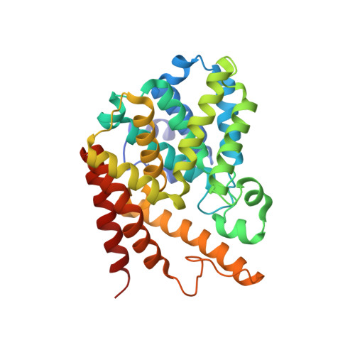
    PDE7 inhibitors regulate pro-inflammatory and immune T-cell functions, and are a potentially novel class of drugs especially useful in the treatment of a wide variety of immune and inflammatory disorders. Starting from our lead family of thioxoquinazolines, we designed, synthesized, and characterized a novel series of thioxoquinazoline derivatives. Many of these compounds showed inhibitory potencies at sub-micromolar levels against the catalytic domain of PDE7A1 and at the micromolar level against PDE4D2. Cell-based studies showed that these compounds not only increased intracellular cAMP levels, but also had interesting anti-inflammatory properties within a therapeutic window. The in silico data predict that these compounds are capable of the crossing the blood-brain barrier. The X-ray crystal structure of the PDE7A1 catalytic domain in complex with compound 15 at a resolution of 2.4 A demonstrated that hydrophobic interactions at the active site pocket are a key feature. This structure, together with molecular modeling, provides insight into the selectivity of the PDE inhibitors and a template for the discovery of new PDE7 or PDE7/PDE4 dual inhibitors.


  • Organizational Affiliation

    Instituto de Química Médica (CSIC), Juan de la Cierva 3, 28006 Madrid, Spain.


Macromolecules
Find similar proteins by:  (by identity cutoff)  |  3D Structure
Entity ID: 1
MoleculeChains Sequence LengthOrganismDetailsImage
High affinity cAMP-specific 3',5'-cyclic phosphodiesterase 7A318Homo sapiensMutation(s): 0 
Gene Names: PDE7APDE7A1
EC: 3.1.4.17
UniProt & NIH Common Fund Data Resources
Find proteins for Q13946 (Homo sapiens)
Explore Q13946 
Go to UniProtKB:  Q13946
PHAROS:  Q13946
GTEx:  ENSG00000205268 
Entity Groups  
Sequence Clusters30% Identity50% Identity70% Identity90% Identity95% Identity100% Identity
UniProt GroupQ13946
Sequence Annotations
Expand
  • Reference Sequence
Small Molecules
Binding Affinity Annotations 
IDSourceBinding Affinity
TC8 PDBBind:  3G3N IC50: 510 (nM) from 1 assay(s)
Binding MOAD:  3G3N IC50: 510 (nM) from 1 assay(s)
Experimental Data & Validation

Experimental Data

  • Method: X-RAY DIFFRACTION
  • Resolution: 2.40 Å
  • R-Value Free: 0.245 
  • R-Value Work: 0.210 
  • R-Value Observed: 0.210 
  • Space Group: P 31 2 1
Unit Cell:
Length ( Å )Angle ( ˚ )
a = 115.447α = 90
b = 115.447β = 90
c = 64.389γ = 120
Software Package:
Software NamePurpose
ADSCdata collection
AMoREphasing
CNSrefinement
HKL-2000data reduction
HKL-2000data scaling

Structure Validation

View Full Validation Report


Ligand Structure Quality Assessment 


Entry History 

Deposition Data

Revision History  (Full details and data files)

  • Version 1.0: 2009-04-28
    Type: Initial release
  • Version 1.1: 2011-07-13
    Changes: Version format compliance
  • Version 1.2: 2024-02-21
    Changes: Data collection, Database references, Derived calculations