


3G0B
Crystal structure of dipeptidyl peptidase IV in complex with TAK-322
- PDB DOI: https://doi.org/10.2210/pdb3G0B/pdb
- Classification: HYDROLASE/HYDROLASE INHIBITOR
- Organism(s): Homo sapiens
- Expression System: Spodoptera frugiperda
- Mutation(s): No
- Deposited: 2009-01-27 Released: 2010-02-16
Experimental Data Snapshot
- Method: X-RAY DIFFRACTION
- Resolution: 2.25 Å
- R-Value Free: 0.242
- R-Value Work: 0.207
- R-Value Observed: 0.209

This is version 2.0 of the entry. See complete history.

Macromolecules
Find similar proteins by:
(by identity cutoff) | 3D Structure
Entity ID: 1 | |||||
|---|---|---|---|---|---|
| Molecule | Chains | Sequence Length | Organism | Details | Image |
| Dipeptidyl peptidase 4 | 740 | Homo sapiens | Mutation(s): 0 Gene Names: ADCP2, CD26, DPP4 EC: 3.4.14.5 | 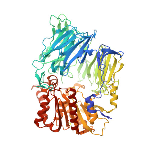 | |
UniProt & NIH Common Fund Data Resources | |||||
Find proteins for P27487 (Homo sapiens) Explore P27487 Go to UniProtKB: P27487 | |||||
PHAROS: P27487 GTEx: ENSG00000197635 | |||||
Entity Groups | |||||
| Sequence Clusters | 30% Identity50% Identity70% Identity90% Identity95% Identity100% Identity | ||||
| UniProt Group | P27487 | ||||
Sequence AnnotationsExpand | |||||
| |||||
Oligosaccharides

Small Molecules
| Ligands 2 Unique | |||||
|---|---|---|---|---|---|
| ID | Chains | Name / Formula / InChI Key | 2D Diagram | 3D Interactions | |
| T22 Query on T22 | AA [auth C], GA [auth D], Q [auth A], V [auth B] | 2-({6-[(3R)-3-aminopiperidin-1-yl]-3-methyl-2,4-dioxo-3,4-dihydropyrimidin-1(2H)-yl}methyl)benzonitrile C18 H21 N5 O2 ZSBOMTDTBDDKMP-OAHLLOKOSA-N |  | ||
| NAG Query on NAG | BA [auth D] CA [auth D] DA [auth D] EA [auth D] FA [auth D] | 2-acetamido-2-deoxy-beta-D-glucopyranose C8 H15 N O6 OVRNDRQMDRJTHS-FMDGEEDCSA-N |  | ||
Experimental Data & Validation
Experimental Data
- Method: X-RAY DIFFRACTION
- Resolution: 2.25 Å
- R-Value Free: 0.242
- R-Value Work: 0.207
- R-Value Observed: 0.209
- Space Group: P 1 21 1
Unit Cell:
| Length ( Å ) | Angle ( ˚ ) |
|---|---|
| a = 121.686 | α = 90 |
| b = 122.398 | β = 114.72 |
| c = 144.01 | γ = 90 |
| Software Name | Purpose |
|---|---|
| ADSC | data collection |
| PHASER | phasing |
| REFMAC | refinement |
| HKL-2000 | data reduction |
| HKL-2000 | data scaling |
Entry History
Deposition Data
- Released Date: 2010-02-16 Deposition Author(s): Zhang, Z., Wallace, M.B., Feng, J., Stafford, J.A., Kaldor, S.W., Shi, L., Skene, R.J., Aertgeerts, K., Lee, B., Jennings, A., Xu, R., Kassel, D., Webb, D.R., Gwaltney, S.L.
Revision History (Full details and data files)
- Version 1.0: 2010-02-16
Type: Initial release - Version 1.1: 2011-07-13
Changes: Advisory, Refinement description, Version format compliance - Version 1.2: 2016-04-06
Changes: Non-polymer description - Version 2.0: 2020-07-29
Type: Remediation
Reason: Carbohydrate remediation
Changes: Advisory, Atomic model, Data collection, Database references, Derived calculations, Structure summary
