3FXB

Crystal structure of the ectoine-binding protein UehA


Experimental Data Snapshot

  • Method: X-RAY DIFFRACTION
  • Resolution: 2.90 Å
  • R-Value Free: 0.262 
  • R-Value Work: 0.218 
  • R-Value Observed: 0.220 

wwPDB Validation   3D Report Full Report


This is version 1.2 of the entry. See complete history


Literature

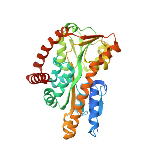
The crystal structure of UehA in complex with ectoine-A comparison with other TRAP-T binding proteins.

Lecher, J.Pittelkow, M.Zobel, S.Bursy, J.Bonig, T.Smits, S.H.Schmitt, L.Bremer, E.

(2009) J Mol Biol 389: 58-73

  • DOI: https://doi.org/10.1016/j.jmb.2009.03.077
  • Primary Citation of Related Structures:  
    3FXB

  • PubMed Abstract: 

    Substrate-binding proteins or extracellular solute receptors (ESRs) are components of both ABC (ATP binding cassette) and TRAP-T (tripartite ATP-independent periplasmic transporter). The TRAP-T system UehABC from Silicibacter pomeroyi DSS-3 imports the compatible solutes ectoine and 5-hydroxyectoine as nutrients. UehA, the ESR of the UehABC operon, binds both ectoine and 5-hydroxyectoine with high affinity (K(d) values of 1.4+/-0.1 and 1.1+/-0.1 microM, respectively) and delivers them to the TRAP-T complex. The crystal structure of UehA in complex with ectoine was determined at 2.9-A resolution and revealed an overall fold common for all ESR proteins from TRAP systems determined so far. A comparison of the recently described structure of TeaA from Halomonas elongata and an ectoine-binding protein (EhuB) from an ABC transporter revealed a conserved ligand binding mode that involves both directed and cation-pi interactions. Furthermore, a comparison with other known TRAP-T ESRs revealed a helix that might act as a selectivity filter imposing restraints on the ESRs that fine-tune ligand recognition and binding and finally might determine the selection of the cognate substrate.


  • Organizational Affiliation

    Institute of Biochemistry, Heinrich-Heine-University Duesseldorf, Germany.


Macromolecules
Find similar proteins by:  (by identity cutoff)  |  3D Structure
Entity ID: 1
MoleculeChains Sequence LengthOrganismDetailsImage
TRAP dicarboxylate transporter, DctP subunit
A, B
326Ruegeria pomeroyi DSS-3Mutation(s): 0 
Gene Names: SPO1147
UniProt
Find proteins for Q5LUA7 (Ruegeria pomeroyi (strain ATCC 700808 / DSM 15171 / DSS-3))
Explore Q5LUA7 
Go to UniProtKB:  Q5LUA7
Entity Groups  
Sequence Clusters30% Identity50% Identity70% Identity90% Identity95% Identity100% Identity
UniProt GroupQ5LUA7
Sequence Annotations
Expand
  • Reference Sequence
Small Molecules
Ligands 1 Unique
IDChains Name / Formula / InChI Key2D Diagram3D Interactions
4CS
Query on 4CS

Download Ideal Coordinates CCD File 
C [auth A],
D [auth B]
(4S)-2-METHYL-1,4,5,6-TETRAHYDROPYRIMIDINE-4-CARBOXYLIC ACID
C6 H10 N2 O2
WQXNXVUDBPYKBA-YFKPBYRVSA-N
Binding Affinity Annotations 
IDSourceBinding Affinity
4CS PDBBind:  3FXB Kd: 1400 (nM) from 1 assay(s)
Experimental Data & Validation

Experimental Data

  • Method: X-RAY DIFFRACTION
  • Resolution: 2.90 Å
  • R-Value Free: 0.262 
  • R-Value Work: 0.218 
  • R-Value Observed: 0.220 
  • Space Group: P 1 21 1
Unit Cell:
Length ( Å )Angle ( ˚ )
a = 63α = 90
b = 61.3β = 102.6
c = 86.3γ = 90
Software Package:
Software NamePurpose
HKL-2000data collection
PHASERphasing
REFMACrefinement
XDSdata reduction
XSCALEdata scaling

Structure Validation

View Full Validation Report


Entry History 

Deposition Data

Revision History  (Full details and data files)

  • Version 1.0: 2009-05-26
    Type: Initial release
  • Version 1.1: 2011-07-13
    Changes: Advisory, Version format compliance
  • Version 1.2: 2023-09-06
    Changes: Data collection, Database references, Derived calculations, Refinement description