3FVG

Crystal structure of the human glutamate receptor, GluR5, ligand-binding core in complex with MSVIII-19 in space group P1


Experimental Data Snapshot

  • Method: X-RAY DIFFRACTION
  • Resolution: 1.50 Å
  • R-Value Free: 0.192 
  • R-Value Work: 0.168 
  • R-Value Observed: 0.169 

wwPDB Validation   3D Report Full Report


Ligand Structure Quality Assessment 


This is version 1.4 of the entry. See complete history


Literature

Binding and Selectivity of the Marine Toxin Neodysiherbaine A and Its Synthetic Analogues to GluK1 and GluK2 Kainate Receptors.

Unno, M.Shinohara, M.Takayama, K.Tanaka, H.Teruya, K.Doh-Ura, K.Sakai, R.Sasaki, M.Ikeda-Saito, M.

(2011) J Mol Biol 413: 667-683

  • DOI: https://doi.org/10.1016/j.jmb.2011.08.043
  • Primary Citation of Related Structures:  
    2ZNS, 2ZNT, 2ZNU, 3FUZ, 3FV1, 3FV2, 3FVG, 3FVK, 3FVN, 3QXM

  • PubMed Abstract: 

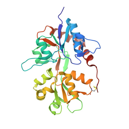
    Dysiherbaine (DH) and neodysiherbaine A (NDH) selectively bind and activate two kainate-type ionotropic glutamate receptors, GluK1 and GluK2. The ligand-binding domains of human GluK1 and GluK2 were crystallized as bound forms with a series of DH analogues including DH, NDH, 8-deoxy-NDH, 9-deoxy-NDH and 8,9-dideoxy-NDH (MSVIII-19), isolated from natural sources or prepared by total synthesis. Since the DH analogues exhibit a wide range of binding affinities and agonist efficacies, it follows that the detailed analysis of crystal structure would provide us with a significant opportunity to elucidate structural factors responsible for selective binding and some aspects of gating efficacy. We found that differences in three amino acids (Thr503, Ser706 and Ser726 in GluK1 and Ala487, Asn690 and Thr710 in GluK2) in the ligand-binding pocket generate differences in the binding modes of NDH to GluK1 and GluK2. Furthermore, deletion of the C(9) hydroxy group in NDH alters the ligand conformation such that it is no longer suited for binding to the GluK1 ligand-binding pocket. In GluK2, NDH pushes and rotates the side chain of Asn690 (substituted for Ser706 in GluK1) and disrupts an interdomain hydrogen bond with Glu409. The present data support the idea that receptor selectivities of DH analogues resulted from the differences in the binding modes of the ligands in GluK1/GluK2 and the steric repulsion of Asn690 in GluK2. All ligands, regardless of agonist efficacy, induced full domain closure. Consequently, ligand efficacy and domain closure did not directly coincide with DH analogues and the kainate receptors.


  • Organizational Affiliation

    Frontier Research Center for Applied Atomic Sciences, Ibaraki University, Tokai, Naka, Ibaraki 319-1106, Japan. unno19@mx.ibaraki.ac.jp


Macromolecules
Find similar proteins by:  (by identity cutoff)  |  3D Structure
Entity ID: 1
MoleculeChains Sequence LengthOrganismDetailsImage
Glutamate receptor, ionotropic kainate 1
A, B
256Homo sapiensMutation(s): 0 
UniProt & NIH Common Fund Data Resources
Find proteins for P39086 (Homo sapiens)
Explore P39086 
Go to UniProtKB:  P39086
PHAROS:  P39086
GTEx:  ENSG00000171189 
Entity Groups  
Sequence Clusters30% Identity50% Identity70% Identity90% Identity95% Identity100% Identity
UniProt GroupP39086
Sequence Annotations
Expand
  • Reference Sequence
Small Molecules
Ligands 3 Unique
IDChains Name / Formula / InChI Key2D Diagram3D Interactions
MS8
Query on MS8

Download Ideal Coordinates CCD File 
F [auth A],
N [auth B]
(2R,3aR,7aR)-2-[(2S)-2-amino-3-hydroxy-3-oxo-propyl]-3,3a,5,6,7,7a-hexahydrofuro[4,5-b]pyran-2-carboxylic acid
C11 H17 N O6
RZIYCCQNKHONBB-PRKAOEEVSA-N
SO4
Query on SO4

Download Ideal Coordinates CCD File 
G [auth A],
H [auth A],
O [auth B],
P [auth B]
SULFATE ION
O4 S
QAOWNCQODCNURD-UHFFFAOYSA-L
GOL
Query on GOL

Download Ideal Coordinates CCD File 
C [auth A]
D [auth A]
E [auth A]
I [auth B]
J [auth B]
C [auth A],
D [auth A],
E [auth A],
I [auth B],
J [auth B],
K [auth B],
L [auth B],
M [auth B]
GLYCEROL
C3 H8 O3
PEDCQBHIVMGVHV-UHFFFAOYSA-N
Binding Affinity Annotations 
IDSourceBinding Affinity
MS8 PDBBind:  3FVG Ki: 128 (nM) from 1 assay(s)
Binding MOAD:  3FVG Ki: 128 (nM) from 1 assay(s)
Experimental Data & Validation

Experimental Data

  • Method: X-RAY DIFFRACTION
  • Resolution: 1.50 Å
  • R-Value Free: 0.192 
  • R-Value Work: 0.168 
  • R-Value Observed: 0.169 
  • Space Group: P 1
Unit Cell:
Length ( Å )Angle ( ˚ )
a = 47.627α = 80.41
b = 50.86β = 84.16
c = 62.931γ = 62.06
Software Package:
Software NamePurpose
CNSrefinement
REFMACrefinement
HKL-2000data reduction
HKL-2000data scaling
CNSphasing

Structure Validation

View Full Validation Report


Ligand Structure Quality Assessment 


Entry History 

Deposition Data

Revision History  (Full details and data files)

  • Version 1.0: 2010-01-19
    Type: Initial release
  • Version 1.1: 2011-07-13
    Changes: Version format compliance
  • Version 1.2: 2011-10-26
    Changes: Database references
  • Version 1.3: 2017-08-16
    Changes: Data collection, Refinement description, Source and taxonomy
  • Version 1.4: 2023-11-01
    Changes: Data collection, Database references, Derived calculations, Refinement description