3FVE

Crystal structure of diaminopimelate epimerase Mycobacterium tuberculosis DapF


Experimental Data Snapshot

  • Method: X-RAY DIFFRACTION
  • Resolution: 2.60 Å
  • R-Value Free: 0.254 
  • R-Value Work: 0.224 
  • R-Value Observed: 0.226 

wwPDB Validation   3D Report Full Report


This is version 1.2 of the entry. See complete history


Literature

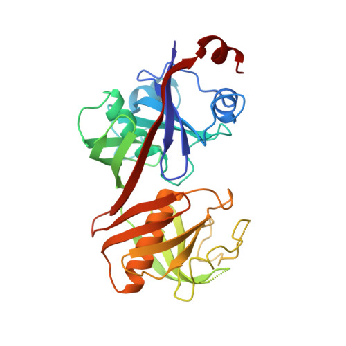
Structure of the diaminopimelate epimerase DapF from Mycobacterium tuberculosis

Usha, V.Dover, L.G.Roper, D.I.Futterer, K.Besra, G.S.

(2009) Acta Crystallogr D Biol Crystallogr 65: 383-387

  • DOI: https://doi.org/10.1107/S0907444909002522
  • Primary Citation of Related Structures:  
    3FVE

  • PubMed Abstract: 

    The meso (or D,L) isomer of diaminopimelic acid (DAP), a precursor of L-lysine, is a key component of the pentapeptide linker in bacterial peptidoglycan. While the peptidoglycan incorporated in the highly complex cell wall of the pathogen Mycobacterium tuberculosis structurally resembles that of Escherichia coli, it is unique in that it can contain penicillin-resistant meso-DAP-->meso-DAP linkages. The interconversion of L,L-DAP and meso-DAP is catalysed by the DAP epimerase DapF, a gene product that is essential in M. tuberculosis. Here, the crystal structure of the ligand-free form of M. tuberculosis DapF (MtDapF) refined to a resolution of 2.6 A is reported. MtDapF shows small if distinct deviations in secondary structure from the two-domain alpha/beta-fold of the known structures of Haemophilus influenzae DapF and Bacillus anthracis DapF, which are in line with its low sequence identity (


  • Organizational Affiliation

    School of Biosciences, University of Birmingham, Edgbaston, Birmingham, England.


Macromolecules
Find similar proteins by:  (by identity cutoff)  |  3D Structure
Entity ID: 1
MoleculeChains Sequence LengthOrganismDetailsImage
Diaminopimelate epimerase290Mycobacterium tuberculosisMutation(s): 0 
Gene Names: dapFMT2798MTCY154.06cRv2726c
EC: 5.1.1.7
UniProt
Find proteins for P9WP19 (Mycobacterium tuberculosis (strain ATCC 25618 / H37Rv))
Explore P9WP19 
Go to UniProtKB:  P9WP19
Entity Groups  
Sequence Clusters30% Identity50% Identity70% Identity90% Identity95% Identity100% Identity
UniProt GroupP9WP19
Sequence Annotations
Expand
  • Reference Sequence
Experimental Data & Validation

Experimental Data

  • Method: X-RAY DIFFRACTION
  • Resolution: 2.60 Å
  • R-Value Free: 0.254 
  • R-Value Work: 0.224 
  • R-Value Observed: 0.226 
  • Space Group: P 65 2 2
Unit Cell:
Length ( Å )Angle ( ˚ )
a = 183.64α = 90
b = 183.64β = 90
c = 45.21γ = 120
Software Package:
Software NamePurpose
PHASERphasing
REFMACrefinement
PDB_EXTRACTdata extraction
ADSCdata collection
XDSdata reduction
SCALAdata scaling

Structure Validation

View Full Validation Report


Entry History 

Deposition Data

Revision History  (Full details and data files)

  • Version 1.0: 2009-01-27
    Type: Initial release
  • Version 1.1: 2011-07-13
    Changes: Advisory, Version format compliance
  • Version 1.2: 2023-09-06
    Changes: Data collection, Database references, Derived calculations, Refinement description