3FS7

Crystal structure of Gallus gallus beta-parvalbumin (avian thymic hormone)


Experimental Data Snapshot

  • Method: X-RAY DIFFRACTION
  • Resolution: 1.95 Å
  • R-Value Free: 0.221 
  • R-Value Work: 0.164 
  • R-Value Observed: 0.166 

wwPDB Validation   3D Report Full Report


This is version 1.3 of the entry. See complete history


Literature

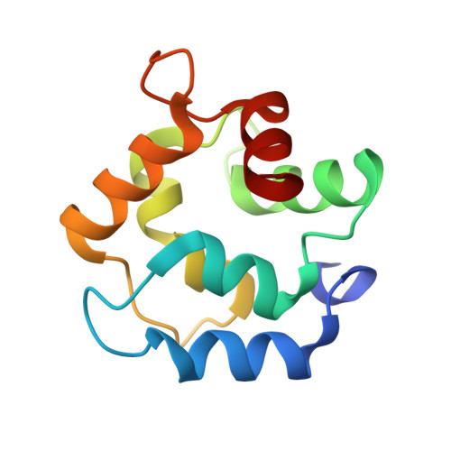
Structure of avian thymic hormone, a high-affinity avian beta-parvalbumin, in the Ca2+-free and Ca2+-bound states.

Schuermann, J.P.Tan, A.Tanner, J.J.Henzl, M.T.

(2010) J Mol Biol 397: 991-1002

  • DOI: https://doi.org/10.1016/j.jmb.2010.02.014
  • Primary Citation of Related Structures:  
    2KQY, 3FS7

  • PubMed Abstract: 

    Originally isolated on the basis of its capacity to stimulate T-cell maturation and proliferation, avian thymic hormone (ATH) is nevertheless a parvalbumin, one of two beta-lineage isoforms expressed in birds. We recently learned that addition of Ca(2+)-free ATH to a solution of 8-anilinonaphthalene-1-sulfonate (ANS) markedly increases ANS emission. This behavior, not observed in the presence of Ca(2+), suggests that apolar surface area buried in the Ca(2+)-bound state becomes solvent accessible upon Ca(2+) removal. In order to elucidate the conformational alterations that accompany Ca(2+) binding, we have obtained the solution structure of the Ca(2+)-free protein using NMR spectroscopy and compared it to the Ca(2+)-loaded protein, solved by X-ray crystallography. Although the metal-ion-binding (CD-EF) domains are largely coincident in the superimposed structures, a major difference is observed in the AB domains. The tight association of helix B with the E and F helices in the Ca(2+)-bound state is lost upon removal of Ca(2+), producing a deep hydrophobic cavity. The B helix also undergoes substantial rotation, exposing the side chains of F24, Y26, F29, and F30 to solvent. Presumably, the increase in ANS emission observed in the presence of unliganded ATH reflects the interaction of these hydrophobic residues with the fluorescent probe. The increased solvent exposure of apolar surface area in the Ca(2+)-free protein is consistent with previously collected scanning calorimetry data, which indicated an unusually low change in heat capacity upon thermal denaturation. The Ca(2+)-free structure also provides added insight into the magnitude of ligation-linked conformational alteration compatible with a high-affinity metal-ion-binding signature. The exposure of substantial apolar surface area suggests the intriguing possibility that ATH could function as a reverse Ca(2+) sensor.


  • Organizational Affiliation

    Northeastern Collaborative Access Team (NE-CAT), Department of Chemistry and Chemical Biology, Cornell University, Ithaca, NY 14853, USA.


Macromolecules
Find similar proteins by:  (by identity cutoff)  |  3D Structure
Entity ID: 1
MoleculeChains Sequence LengthOrganismDetailsImage
Parvalbumin, thymic
A, B, C, D, E
A, B, C, D, E, F, G, H
109Gallus gallusMutation(s): 0 
UniProt
Find proteins for P19753 (Gallus gallus)
Explore P19753 
Go to UniProtKB:  P19753
Entity Groups  
Sequence Clusters30% Identity50% Identity70% Identity90% Identity95% Identity100% Identity
UniProt GroupP19753
Sequence Annotations
Expand
  • Reference Sequence
Small Molecules
Ligands 3 Unique
IDChains Name / Formula / InChI Key2D Diagram3D Interactions
SO4
Query on SO4

Download Ideal Coordinates CCD File 
BA [auth G],
EA [auth H],
K [auth A],
N [auth B]
SULFATE ION
O4 S
QAOWNCQODCNURD-UHFFFAOYSA-L
GOL
Query on GOL

Download Ideal Coordinates CCD File 
Q [auth C],
T [auth D],
Y [auth F]
GLYCEROL
C3 H8 O3
PEDCQBHIVMGVHV-UHFFFAOYSA-N
CA
Query on CA

Download Ideal Coordinates CCD File 
AA [auth G]
CA [auth H]
DA [auth H]
I [auth A]
J [auth A]
AA [auth G],
CA [auth H],
DA [auth H],
I [auth A],
J [auth A],
L [auth B],
M [auth B],
O [auth C],
P [auth C],
R [auth D],
S [auth D],
U [auth E],
V [auth E],
W [auth F],
X [auth F],
Z [auth G]
CALCIUM ION
Ca
BHPQYMZQTOCNFJ-UHFFFAOYSA-N
Experimental Data & Validation

Experimental Data

  • Method: X-RAY DIFFRACTION
  • Resolution: 1.95 Å
  • R-Value Free: 0.221 
  • R-Value Work: 0.164 
  • R-Value Observed: 0.166 
  • Space Group: P 1
Unit Cell:
Length ( Å )Angle ( ˚ )
a = 47.644α = 87.42
b = 51.56β = 85.42
c = 67.633γ = 87.42
Software Package:
Software NamePurpose
PHENIXrefinement
PDB_EXTRACTdata extraction
HKL-2000data collection
HKL-2000data reduction
HKL-2000data scaling
MOLREPphasing
REFMACrefinement

Structure Validation

View Full Validation Report


Entry History 

Deposition Data

Revision History  (Full details and data files)

  • Version 1.0: 2010-01-19
    Type: Initial release
  • Version 1.1: 2011-07-13
    Changes: Version format compliance
  • Version 1.2: 2019-07-24
    Changes: Data collection, Refinement description
  • Version 1.3: 2023-09-06
    Changes: Data collection, Database references, Derived calculations, Refinement description