3FKQ

Crystal structure of NtrC-like two-domain protein (RER070207001320) from Eubacterium rectale at 2.10 A resolution


Experimental Data Snapshot

  • Method: X-RAY DIFFRACTION
  • Resolution: 2.10 Å
  • R-Value Free: 0.218 
  • R-Value Work: 0.190 
  • R-Value Observed: 0.191 

wwPDB Validation   3D Report Full Report


Ligand Structure Quality Assessment 


This is version 1.4 of the entry. See complete history


Literature

Structure of the pilus assembly protein TadZ from Eubacterium rectale: implications for polar localization.

Xu, Q.Christen, B.Chiu, H.J.Jaroszewski, L.Klock, H.E.Knuth, M.W.Miller, M.D.Elsliger, M.A.Deacon, A.M.Godzik, A.Lesley, S.A.Figurski, D.H.Shapiro, L.Wilson, I.A.

(2012) Mol Microbiol 83: 712-727

  • DOI: https://doi.org/10.1111/j.1365-2958.2011.07954.x
  • Primary Citation of Related Structures:  
    3FKQ

  • PubMed Abstract: 

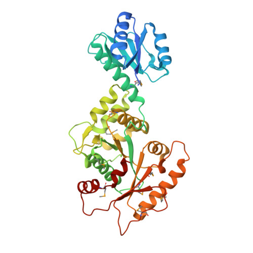
    The tad (tight adherence) locus encodes a protein translocation system that produces a novel variant of type IV pili. The pilus assembly protein TadZ (called CpaE in Caulobacter crescentus) is ubiquitous in tad loci, but is absent in other type IV pilus biogenesis systems. The crystal structure of TadZ from Eubacterium rectale (ErTadZ), in complex with ATP and Mg(2+) , was determined to 2.1 Å resolution. ErTadZ contains an atypical ATPase domain with a variant of a deviant Walker-A motif that retains ATP binding capacity while displaying only low intrinsic ATPase activity. The bound ATP plays an important role in dimerization of ErTadZ. The N-terminal atypical receiver domain resembles the canonical receiver domain of response regulators, but has a degenerate, stripped-down 'active site'. Homology modelling of the N-terminal atypical receiver domain of CpaE indicates that it has a conserved protein-protein binding surface similar to that of the polar localization module of the social mobility protein FrzS, suggesting a similar function. Our structural results also suggest that TadZ localizes to the pole through the atypical receiver domain during an early stage of pili biogenesis, and functions as a hub for recruiting other pili components, thus providing insights into the Tad pilus assembly process.


  • Organizational Affiliation

    Joint Center for Structural Genomics.


Macromolecules
Find similar proteins by:  (by identity cutoff)  |  3D Structure
Entity ID: 1
MoleculeChains Sequence LengthOrganismDetailsImage
NtrC-like two-domain protein373Agathobacter rectalisMutation(s): 0 
Gene Names: RER070207001320
UniProt
Find proteins for D0VX10 (Agathobacter rectalis)
Explore D0VX10 
Go to UniProtKB:  D0VX10
Entity Groups  
Sequence Clusters30% Identity50% Identity70% Identity90% Identity95% Identity100% Identity
UniProt GroupD0VX10
Sequence Annotations
Expand
  • Reference Sequence
Small Molecules
Ligands 6 Unique
IDChains Name / Formula / InChI Key2D Diagram3D Interactions
ATP
Query on ATP

Download Ideal Coordinates CCD File 
B [auth A]ADENOSINE-5'-TRIPHOSPHATE
C10 H16 N5 O13 P3
ZKHQWZAMYRWXGA-KQYNXXCUSA-N
2PE
Query on 2PE

Download Ideal Coordinates CCD File 
E [auth A],
F [auth A]
NONAETHYLENE GLYCOL
C18 H38 O10
YZUUTMGDONTGTN-UHFFFAOYSA-N
SO4
Query on SO4

Download Ideal Coordinates CCD File 
I [auth A]SULFATE ION
O4 S
QAOWNCQODCNURD-UHFFFAOYSA-L
GOL
Query on GOL

Download Ideal Coordinates CCD File 
G [auth A],
H [auth A]
GLYCEROL
C3 H8 O3
PEDCQBHIVMGVHV-UHFFFAOYSA-N
CL
Query on CL

Download Ideal Coordinates CCD File 
D [auth A]CHLORIDE ION
Cl
VEXZGXHMUGYJMC-UHFFFAOYSA-M
MG
Query on MG

Download Ideal Coordinates CCD File 
C [auth A]MAGNESIUM ION
Mg
JLVVSXFLKOJNIY-UHFFFAOYSA-N
Modified Residues  1 Unique
IDChains TypeFormula2D DiagramParent
MSE
Query on MSE
A
L-PEPTIDE LINKINGC5 H11 N O2 SeMET
Experimental Data & Validation

Experimental Data

Unit Cell:
Length ( Å )Angle ( ˚ )
a = 121.506α = 90
b = 79.495β = 96.16
c = 55.022γ = 90
Software Package:
Software NamePurpose
REFMACrefinement
PHENIXrefinement
SHELXphasing
MolProbitymodel building
SCALAdata scaling
PDB_EXTRACTdata extraction
MOSFLMdata reduction
SHELXDphasing
autoSHARPphasing

Structure Validation

View Full Validation Report


Ligand Structure Quality Assessment 


Entry History 

Deposition Data

Revision History  (Full details and data files)

  • Version 1.0: 2009-01-06
    Type: Initial release
  • Version 1.1: 2011-07-13
    Changes: Advisory, Version format compliance
  • Version 1.2: 2012-03-14
    Changes: Database references
  • Version 1.3: 2017-10-25
    Changes: Author supporting evidence, Refinement description
  • Version 1.4: 2019-07-24
    Changes: Data collection, Database references, Derived calculations, Refinement description