Structural basis for autoinhibition and activation of Auto, a virulence-associated peptidoglycan hydrolase of Listeria monocytogenes.
Bublitz, M., Polle, L., Holland, C., Heinz, D.W., Nimtz, M., Schubert, W.D.(2009) Mol Microbiol 71: 1509-1522
- PubMed: 19210622
- DOI: https://doi.org/10.1111/j.1365-2958.2009.06619.x
- Primary Citation of Related Structures:
3FI7 - PubMed Abstract:
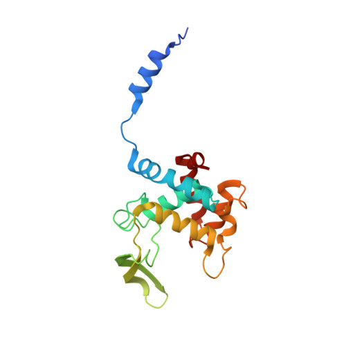
During a bacterial infection, each successive step is orchestrated by a dedicated set of virulence factors. In Gram-positive bacteria, the presentation or release of such factors is crucially dependent on the continual remodelling of the cell wall. We have investigated the autolysin or peptidoglycan hydrolase Auto (Lmo1076) from the human pathogen Listeria monocytogenes to structurally and biochemically underpin its role in host cell invasion. We demonstrate that Auto is an N-acetylglucosaminidase, that it is autoinhibited when newly secreted but activated by proteolytic cleavage, that it has an acidic pH optimum and that it preferentially cleaves acetylated over de-acetylated peptidoglycan. The crystal structure of Auto, the first for glycoside hydrolase family 73, and the first for a listerial autolysin, indicates that autoinhibition is due to an N-terminal alpha-helix unique to Auto that physically blocks the substrate-binding cleft. We identify Glu122 and Glu156 as the two catalytically essential carboxylate groups. The physical properties of Auto as well as its localization to lipoteichoic acid by its four C-terminal GW modules imply cell wall degradation by Auto to be highly co-ordinated. Its spatio-temporally controlled activation and localized activity in an acidified environment indicate that it facilitates remodelling of the cell wall and may be involved in co-ordinating the release of virulence factors at specific stages of an infection.
Organizational Affiliation:
Molecular Host Pathogen Interactions, Division of Structural Biology, Helmholtz Centre for Infection Research, Inhoffenstr, Braunschweig, Germany.