3FHG

Crystal structure of Sulfolobus solfataricus 8-oxoguanine DNA glycosylase (SsOgg)


Experimental Data Snapshot

  • Method: X-RAY DIFFRACTION
  • Resolution: 1.90 Å
  • R-Value Free: 0.248 
  • R-Value Work: 0.207 
  • R-Value Observed: 0.226 

wwPDB Validation   3D Report Full Report


This is version 1.4 of the entry. See complete history


Literature

Crystal structures of two archaeal 8-oxoguanine DNA glycosylases provide structural insight into guanine/8-oxoguanine distinction.

Faucher, F.Duclos, S.Bandaru, V.Wallace, S.S.Doublie, S.

(2009) Structure 17: 703-712

  • DOI: https://doi.org/10.1016/j.str.2009.03.007
  • Primary Citation of Related Structures:  
    3FHF, 3FHG

  • PubMed Abstract: 

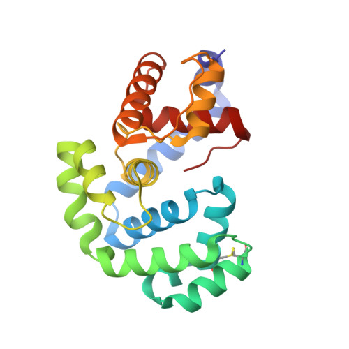
    Among the four DNA bases, guanine is particularly vulnerable to oxidative damage and the most common oxidative product, 7,8-dihydro-8-oxoguanine (8-oxoG), is the most prevalent lesion observed in DNA molecules. Fortunately, 8-oxoG is recognized and excised by the 8-oxoguanine DNA glycosylase (Ogg) of the base excision repair pathway. Ogg enzymes are divided into three separate families, namely, Ogg1, Ogg2, and archaeal GO glycosylase (AGOG). To date, structures of members of both Ogg1 and AGOG families are known but no structural information is available for members of Ogg2. Here we describe the first crystal structures of two archaeal Ogg2: Methanocaldococcus janischii Ogg and Sulfolobus solfataricus Ogg. A structural comparison with OGG1 and AGOG suggested that the C-terminal lysine of Ogg2 may play a key role in discriminating between guanine and 8-oxoG. This prediction was substantiated by measuring the glycosylase/lyase activity of a C-terminal deletion mutant of MjaOgg.


  • Organizational Affiliation

    Department of Microbiology and Molecular Genetics, The Markey Center for Molecular Genetics, University of Vermont, Stafford Hall, Burlington, VT 05405-0068, USA.


Macromolecules
Find similar proteins by:  (by identity cutoff)  |  3D Structure
Entity ID: 1
MoleculeChains Sequence LengthOrganismDetailsImage
N-glycosylase/DNA lyase207Saccharolobus solfataricusMutation(s): 1 
Gene Names: 1451601MJ0724oggSSO0904
EC: 3.2.2 (PDB Primary Data), 4.2.99.18 (PDB Primary Data)
UniProt
Find proteins for Q97ZK2 (Saccharolobus solfataricus (strain ATCC 35092 / DSM 1617 / JCM 11322 / P2))
Explore Q97ZK2 
Go to UniProtKB:  Q97ZK2
Entity Groups  
Sequence Clusters30% Identity50% Identity70% Identity90% Identity95% Identity100% Identity
UniProt GroupQ97ZK2
Sequence Annotations
Expand
  • Reference Sequence
Small Molecules
Ligands 2 Unique
IDChains Name / Formula / InChI Key2D Diagram3D Interactions
SO4
Query on SO4

Download Ideal Coordinates CCD File 
G [auth A]
H [auth A]
I [auth A]
J [auth A]
K [auth A]
G [auth A],
H [auth A],
I [auth A],
J [auth A],
K [auth A],
L [auth A],
M [auth A],
N [auth A],
O [auth A],
P [auth A]
SULFATE ION
O4 S
QAOWNCQODCNURD-UHFFFAOYSA-L
GOL
Query on GOL

Download Ideal Coordinates CCD File 
B [auth A],
C [auth A],
D [auth A],
E [auth A],
F [auth A]
GLYCEROL
C3 H8 O3
PEDCQBHIVMGVHV-UHFFFAOYSA-N
Experimental Data & Validation

Experimental Data

  • Method: X-RAY DIFFRACTION
  • Resolution: 1.90 Å
  • R-Value Free: 0.248 
  • R-Value Work: 0.207 
  • R-Value Observed: 0.226 
  • Space Group: P 6
Unit Cell:
Length ( Å )Angle ( ˚ )
a = 110.96α = 90
b = 110.96β = 90
c = 34.41γ = 120
Software Package:
Software NamePurpose
XSCALEdata processing
CNSrefinement
PDB_EXTRACTdata extraction
Blu-Icedata collection
XDSdata reduction
XSCALEdata scaling
MOLREPphasing

Structure Validation

View Full Validation Report


Entry History 

Deposition Data

Revision History  (Full details and data files)

  • Version 1.0: 2009-05-19
    Type: Initial release
  • Version 1.1: 2011-07-13
    Changes: Non-polymer description, Version format compliance
  • Version 1.2: 2012-03-21
    Changes: Database references
  • Version 1.3: 2021-10-20
    Changes: Database references, Derived calculations
  • Version 1.4: 2023-09-06
    Changes: Data collection, Refinement description