3FGP

2.05 a Crystal Structure of CysM from Mycobacterium Tuberculosis - Open and Closed Conformations


Experimental Data Snapshot

  • Method: X-RAY DIFFRACTION
  • Resolution: 2.05 Å
  • R-Value Free: 0.247 
  • R-Value Work: 0.198 
  • R-Value Observed: 0.200 

wwPDB Validation   3D Report Full Report


This is version 1.4 of the entry. See complete history


Literature

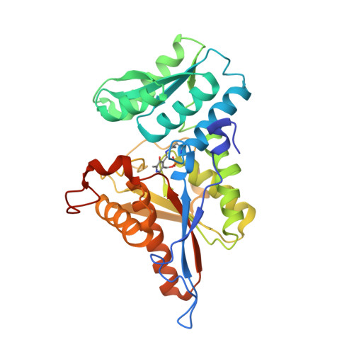
The C-terminal of CysM from Mycobacterium tuberculosis protects the aminoacrylate intermediate and is involved in sulfur donor selectivity

Agren, D.Schnell, R.Schneider, G.

(2009) FEBS Lett 583: 330-336

  • DOI: https://doi.org/10.1016/j.febslet.2008.12.019
  • Primary Citation of Related Structures:  
    3FGP

  • PubMed Abstract: 

    A new crystal structure of the dimeric cysteine synthase CysM from Mycobacterium tuberculosis reveals an open and a closed conformation of the enzyme. In the closed conformation the five carboxy-terminal amino acid residues are inserted into the active site cleft. Removal of this segment results in a decreased lifetime of the alpha-aminoacrylate reaction intermediate, an increased sensitivity to oxidants such as hydrogen peroxide, and loss of substrate selectivity with respect to the sulfur carrier thiocarboxylated CysO. These results highlight features of CysM that might be of particular importance for cysteine biosynthesis under oxidative stress in M. tuberculosis.


  • Organizational Affiliation

    Department of Medical Biochemistry and Biophysics, Karolinska Institutet, Scheeles vg 2, S-171 77 Stockholm, Sweden.


Macromolecules
Find similar proteins by:  (by identity cutoff)  |  3D Structure
Entity ID: 1
MoleculeChains Sequence LengthOrganismDetailsImage
Cysteine synthase B
A, B
326Mycobacterium tuberculosisMutation(s): 1 
Gene Names: cysM (Rv1336)
EC: 2.5.1.65
UniProt
Find proteins for P9WP53 (Mycobacterium tuberculosis (strain ATCC 25618 / H37Rv))
Explore P9WP53 
Go to UniProtKB:  P9WP53
Entity Groups  
Sequence Clusters30% Identity50% Identity70% Identity90% Identity95% Identity100% Identity
UniProt GroupP9WP53
Sequence Annotations
Expand
  • Reference Sequence
Small Molecules
Modified Residues  1 Unique
IDChains TypeFormula2D DiagramParent
LLP
Query on LLP
A, B
L-PEPTIDE LINKINGC14 H22 N3 O7 PLYS
Experimental Data & Validation

Experimental Data

  • Method: X-RAY DIFFRACTION
  • Resolution: 2.05 Å
  • R-Value Free: 0.247 
  • R-Value Work: 0.198 
  • R-Value Observed: 0.200 
  • Space Group: P 21 21 21
Unit Cell:
Length ( Å )Angle ( ˚ )
a = 74.933α = 90
b = 89.142β = 90
c = 99.382γ = 90
Software Package:
Software NamePurpose
DNAdata collection
MOLREPphasing
REFMACrefinement
MOSFLMdata reduction
SCALAdata scaling

Structure Validation

View Full Validation Report


Entry History 

Deposition Data

Revision History  (Full details and data files)

  • Version 1.0: 2008-12-23
    Type: Initial release
  • Version 1.1: 2011-07-13
    Changes: Version format compliance
  • Version 1.2: 2021-11-10
    Changes: Database references, Derived calculations
  • Version 1.3: 2023-11-01
    Changes: Data collection, Refinement description
  • Version 1.4: 2023-11-22
    Changes: Data collection