


Crystal structure of PAS domain of RHA05790
Chang, C., Xu, X., Cui, H., Savchenko, A., Edwards, A., Joachimiak, A.To be published.
Experimental Data Snapshot
Entity ID: 1 | |||||
|---|---|---|---|---|---|
| Molecule | Chains | Sequence Length | Organism | Details | Image |
| uncharacterized protein RHA05790 | 118 | Rhodococcus jostii RHA1 | Mutation(s): 0 Gene Names: RHA1_ro04714 | 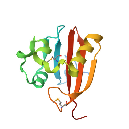 | |
UniProt | |||||
Find proteins for Q0S7I6 (Rhodococcus jostii (strain RHA1)) Explore Q0S7I6 Go to UniProtKB: Q0S7I6 | |||||
Entity Groups | |||||
| Sequence Clusters | 30% Identity50% Identity70% Identity90% Identity95% Identity100% Identity | ||||
| UniProt Group | Q0S7I6 | ||||
Sequence AnnotationsExpand | |||||
| |||||
| Ligands 1 Unique | |||||
|---|---|---|---|---|---|
| ID | Chains | Name / Formula / InChI Key | 2D Diagram | 3D Interactions | |
| 3PB Query on 3PB | G [auth A] H [auth B] I [auth C] J [auth D] K [auth E] | (3R)-3-(phosphonooxy)butanoic acid C4 H9 O6 P CEWUUYVGYVJSGW-GSVOUGTGSA-N |  | ||
| Modified Residues 1 Unique | |||||
|---|---|---|---|---|---|
| ID | Chains | Type | Formula | 2D Diagram | Parent |
| MSE Query on MSE | A, B, C, D, E A, B, C, D, E, F | L-PEPTIDE LINKING | C5 H11 N O2 Se |  | MET |
| Length ( Å ) | Angle ( ˚ ) |
|---|---|
| a = 149.695 | α = 90 |
| b = 55.889 | β = 132.61 |
| c = 101.856 | γ = 90 |
| Software Name | Purpose |
|---|---|
| SBC-Collect | data collection |
| HKL-3000 | phasing |
| MLPHARE | phasing |
| DM | model building |
| SHELXD | phasing |
| RESOLVE | model building |
| Coot | model building |
| REFMAC | refinement |
| HKL-3000 | data reduction |
| HKL-3000 | data scaling |
| DM | phasing |
| RESOLVE | phasing |
RCSB PDB (citation) is hosted by
RCSB PDB is a member of the

