Protein Binding and the Electronic Properties of Iron(II) Complexes: An Electrochemical and Optical Investigation of Outer Sphere Effects.
Barker, K.D., Eckermann, A.L., Sazinsky, M.H., Hartings, M.R., Abajian, C., Georganopoulou, D., Ratner, M.A., Rosenzweig, A.C., Meade, T.J.(2009) Bioconjug Chem 20: 1930-1939
- PubMed: 19788194
- DOI: https://doi.org/10.1021/bc900270a
- Primary Citation of Related Structures:
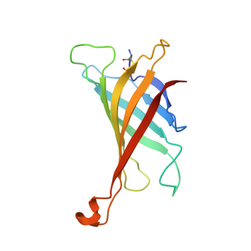
3FDC - PubMed Abstract:
Metalloenzymes and electron transfer proteins influence the electrochemical properties of metal cofactors by controlling the second-sphere environment of the protein active site. Properties that tune this environment include the dielectric constant, templated charge structure, van der Waals interactions, and hydrogen bonds. By systematically varying the binding of a redox-active ligand with a protein, we can evaluate how these noncovalent interactions alter the electronic structure of the bound metal complex. For this study, we employ the well-characterized avidin-biotin conjugate as the protein-ligand system, and have synthesized solvatochromic biotinylated and desthiobiotinylated iron(II) bipyridine tetracyano complexes ([Fe(BMB)(CN)(4)](2-) (1) and [Fe(DMB)(CN)(4)](2-) (2)). The binding affinities of 1 and 2 with avidin are 3.5 × 10(7) M(-1) and 1.5 × 10(6) M(-1), respectively. The redox potentials of 1 and 2 (333 mV and 330 mV) shift to 193 mV and 203 mV vs Ag/AgCl when the complex is bound to avidin and adsorbed to a monolayer-coated gold electrode. Upon binding to avidin, the MLCT1 band red-shifts 20 nm for 1 and 10 nm for 2. Similarly, the MLCT2 band for 1 red-shifts 7 nm and the band for 2 red-shifts 6 nm. For comparison, the electronic properties of 1 and 2 were investigated in organic solvents, and similar shifts in the MLCT bands and redox potentials were observed. An X-ray crystal structure of 1 bound to avidin was obtained, and molecular dynamics simulations were performed to analyze the protein environment of the protein-bound transition metal complexes. Our studies demonstrate that changes in the binding affinity of a ligand-receptor pair influence the outer-sphere coordination of the ligand, which in turn affects the electronic properties of the bound complex.
Organizational Affiliation:
Department of Chemistry, Northwestern University, 2145 Sheridan Road, Evanston, IL 60208, USA.