3FD2

Crystal structure of mMsoI/DNA complex with calcium


Experimental Data Snapshot

  • Method: X-RAY DIFFRACTION
  • Resolution: 2.69 Å
  • R-Value Free: 0.281 
  • R-Value Work: 0.211 
  • R-Value Observed: 0.215 

wwPDB Validation   3D Report Full Report


This is version 1.4 of the entry. See complete history


Literature

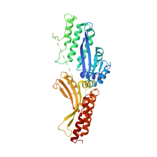
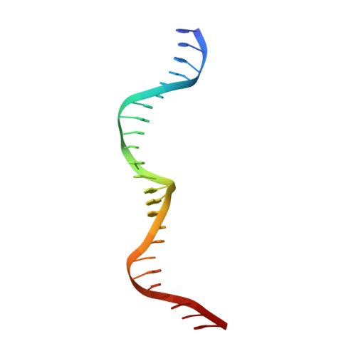
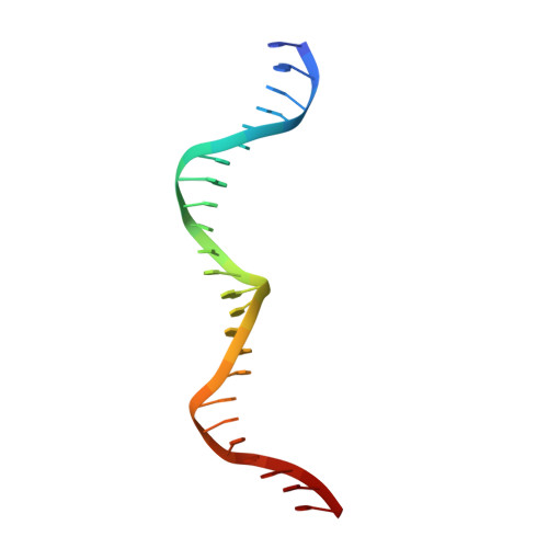
Generation of single-chain LAGLIDADG homing endonucleases from native homodimeric precursor proteins.

Li, H.Pellenz, S.Ulge, U.Stoddard, B.L.Monnat, R.J.

(2009) Nucleic Acids Res 37: 1650-1662

  • DOI: https://doi.org/10.1093/nar/gkp004
  • Primary Citation of Related Structures:  
    3FD2

  • PubMed Abstract: 

    Homing endonucleases (HEs) cut long DNA target sites with high specificity to initiate and target the lateral transfer of mobile introns or inteins. This high site specificity of HEs makes them attractive reagents for gene targeting to promote DNA modification or repair. We have generated several hundred catalytically active, monomerized versions of the well-characterized homodimeric I-CreI and I-MsoI LAGLIDADG family homing endonuclease (LHE) proteins. Representative monomerized I-CreI and I-MsoI proteins (collectively termed mCreIs or mMsoIs) were characterized in detail by using a combination of biochemical, biophysical and structural approaches. We also demonstrated that both mCreI and mMsoI proteins can promote cleavage-dependent recombination in human cells. The use of single chain LHEs should simplify gene modification and targeting by requiring the expression of a single small protein in cells, rather than the coordinate expression of two separate protein coding genes as is required when using engineered heterodimeric zinc finger or homing endonuclease proteins.


  • Organizational Affiliation

    Department of Pathology, University of Washington, Box 357705, Seattle, WA 98195, USA.


Macromolecules

Find similar proteins by:  (by identity cutoff)  |  3D Structure
Entity ID: 1
MoleculeChains Sequence LengthOrganismDetailsImage
Site-specific DNA endonuclease I-MsoI373Monomastix sp. OKE-1Mutation(s): 0 
Gene Names: orf170the chloroplast large subunit rDNA intron gene
EC: 3.1
UniProt
Find proteins for C0JWR6 (Monomastix sp. (strain OKE-1))
Explore C0JWR6 
Go to UniProtKB:  C0JWR6
Entity Groups  
Sequence Clusters30% Identity50% Identity70% Identity90% Identity95% Identity100% Identity
UniProt GroupC0JWR6
Sequence Annotations
Expand
  • Reference Sequence

Find similar nucleic acids by:  Sequence   |   3D Structure  

Entity ID: 2
MoleculeChains LengthOrganismImage
5'-D(*GP*CP*AP*GP*AP*AP*CP*GP*TP*CP*GP*TP*GP*AP*GP*AP*CP*AP*GP*TP*TP*CP*CP*G)-3'24synthetic construct
Sequence Annotations
Expand
  • Reference Sequence

Find similar nucleic acids by:  Sequence   |   3D Structure  

Entity ID: 3
MoleculeChains LengthOrganismImage
5'-D(*CP*GP*GP*AP*AP*CP*TP*GP*TP*CP*TP*CP*AP*CP*GP*AP*CP*GP*TP*TP*CP*TP*GP*C)-3'24synthetic construct
Sequence Annotations
Expand
  • Reference Sequence
Experimental Data & Validation

Experimental Data

  • Method: X-RAY DIFFRACTION
  • Resolution: 2.69 Å
  • R-Value Free: 0.281 
  • R-Value Work: 0.211 
  • R-Value Observed: 0.215 
  • Space Group: P 1
Unit Cell:
Length ( Å )Angle ( ˚ )
a = 41.932α = 107.21
b = 41.933β = 95.43
c = 70.96γ = 109.44
Software Package:
Software NamePurpose
HKL-2000data collection
PHASESphasing
REFMACrefinement
HKL-2000data reduction
HKL-2000data scaling

Structure Validation

View Full Validation Report


Entry History 

Deposition Data

Revision History  (Full details and data files)

  • Version 1.0: 2009-06-30
    Type: Initial release
  • Version 1.1: 2011-07-13
    Changes: Advisory, Refinement description, Version format compliance
  • Version 1.2: 2014-04-09
    Changes: Source and taxonomy
  • Version 1.3: 2017-06-28
    Changes: Database references, Derived calculations, Source and taxonomy, Structure summary
  • Version 1.4: 2023-09-06
    Changes: Data collection, Database references, Derived calculations, Refinement description