3FBV

Crystal structure of the oligomer formed by the kinase-ribonuclease domain of Ire1


Experimental Data Snapshot

  • Method: X-RAY DIFFRACTION
  • Resolution: 3.20 Å
  • R-Value Free: 0.283 
  • R-Value Work: 0.235 
  • R-Value Observed: 0.237 

wwPDB Validation   3D Report Full Report


Ligand Structure Quality Assessment 


This is version 1.3 of the entry. See complete history


Literature

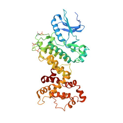
The unfolded protein response signals through high-order assembly of Ire1.

Korennykh, A.V.Egea, P.F.Korostelev, A.A.Finer-Moore, J.Zhang, C.Shokat, K.M.Stroud, R.M.Walter, P.

(2009) Nature 457: 687-693

  • DOI: https://doi.org/10.1038/nature07661
  • Primary Citation of Related Structures:  
    3FBV

  • PubMed Abstract: 

    Aberrant folding of proteins in the endoplasmic reticulum activates the bifunctional transmembrane kinase/endoribonuclease Ire1. Ire1 excises an intron from HAC1 messenger RNA in yeasts and Xbp1 messenger RNA in metozoans encoding homologous transcription factors. This non-conventional mRNA splicing event initiates the unfolded protein response, a transcriptional program that relieves the endoplasmic reticulum stress. Here we show that oligomerization is central to Ire1 function and is an intrinsic attribute of its cytosolic domains. We obtained the 3.2-A crystal structure of the oligomer of the Ire1 cytosolic domains in complex with a kinase inhibitor that acts as a potent activator of the Ire1 RNase. The structure reveals a rod-shaped assembly that has no known precedence among kinases. This assembly positions the kinase domain for trans-autophosphorylation, orders the RNase domain, and creates an interaction surface for binding of the mRNA substrate. Activation of Ire1 through oligomerization expands the mechanistic repertoire of kinase-based signalling receptors.


  • Organizational Affiliation

    Department of Biochemistry and Biophysics, University of California at San Francisco, San Francisco, California 94158, USA. alexei.korennykh@ucsf.edu


Macromolecules
Find similar proteins by:  (by identity cutoff)  |  3D Structure
Entity ID: 1
MoleculeChains Sequence LengthOrganismDetailsImage
Serine/threonine-protein kinase/endoribonuclease IRE1
A, B, C, D, E
A, B, C, D, E, F, G, H, I, J, K, L, M, N
448Saccharomyces cerevisiae S288CMutation(s): 0 
Gene Names: IRE1ERN1
EC: 2.7.11.1 (PDB Primary Data), 3.1.26 (PDB Primary Data)
UniProt
Find proteins for P32361 (Saccharomyces cerevisiae (strain ATCC 204508 / S288c))
Explore P32361 
Go to UniProtKB:  P32361
Entity Groups  
Sequence Clusters30% Identity50% Identity70% Identity90% Identity95% Identity100% Identity
UniProt GroupP32361
Sequence Annotations
Expand
  • Reference Sequence
Small Molecules
Ligands 1 Unique
IDChains Name / Formula / InChI Key2D Diagram3D Interactions
APJ
Query on APJ

Download Ideal Coordinates CCD File 
AA [auth M]
BA [auth N]
O [auth A]
P [auth B]
Q [auth C]
AA [auth M],
BA [auth N],
O [auth A],
P [auth B],
Q [auth C],
R [auth D],
S [auth E],
T [auth F],
U [auth G],
V [auth H],
W [auth I],
X [auth J],
Y [auth K],
Z [auth L]
N~2~-1H-benzimidazol-5-yl-N~4~-(3-cyclopropyl-1H-pyrazol-5-yl)pyrimidine-2,4-diamine
C17 H16 N8
WJNBSTLIALIIEW-UHFFFAOYSA-N
Modified Residues  2 Unique
IDChains TypeFormula2D DiagramParent
SEP
Query on SEP
A, B, C, D, E
A, B, C, D, E, F, G, H, I, J, K, L, M, N
L-PEPTIDE LINKINGC3 H8 N O6 PSER
TPO
Query on TPO
A, B, C, D, E
A, B, C, D, E, F, G, H, I, J, K, L, M, N
L-PEPTIDE LINKINGC4 H10 N O6 PTHR
Experimental Data & Validation

Experimental Data

  • Method: X-RAY DIFFRACTION
  • Resolution: 3.20 Å
  • R-Value Free: 0.283 
  • R-Value Work: 0.235 
  • R-Value Observed: 0.237 
  • Space Group: P 21 21 2
Unit Cell:
Length ( Å )Angle ( ˚ )
a = 156.82α = 90
b = 163.47β = 90
c = 292.83γ = 90
Software Package:
Software NamePurpose
ELVESrefinement
PHASERphasing
PHENIXrefinement
XDSdata reduction
SCALAdata scaling

Structure Validation

View Full Validation Report


Ligand Structure Quality Assessment 


Entry History 

Deposition Data

Revision History  (Full details and data files)

  • Version 1.0: 2008-12-16
    Type: Initial release
  • Version 1.1: 2011-07-13
    Changes: Source and taxonomy, Version format compliance
  • Version 1.2: 2017-11-01
    Changes: Refinement description
  • Version 1.3: 2023-09-06
    Changes: Data collection, Database references, Derived calculations, Refinement description