3FAT

X-ray structure of iGluR4 flip ligand-binding core (S1S2) in complex with (S)-AMPA at 1.90A resolution


Experimental Data Snapshot

  • Method: X-RAY DIFFRACTION
  • Resolution: 1.90 Å
  • R-Value Free: 0.216 
  • R-Value Work: 0.175 
  • R-Value Observed: 0.177 

wwPDB Validation   3D Report Full Report


Ligand Structure Quality Assessment 


This is version 1.3 of the entry. See complete history


Literature

Molecular mechanism of agonist recognition by the ligand-binding core of the ionotropic glutamate receptor 4

Kasper, C.Frydenvang, K.Naur, P.Gajhede, M.Pickering, D.S.Kastrup, J.S.

(2008) FEBS Lett 582: 4089-4094

  • DOI: https://doi.org/10.1016/j.febslet.2008.11.005
  • Primary Citation of Related Structures:  
    3FAS, 3FAT

  • PubMed Abstract: 

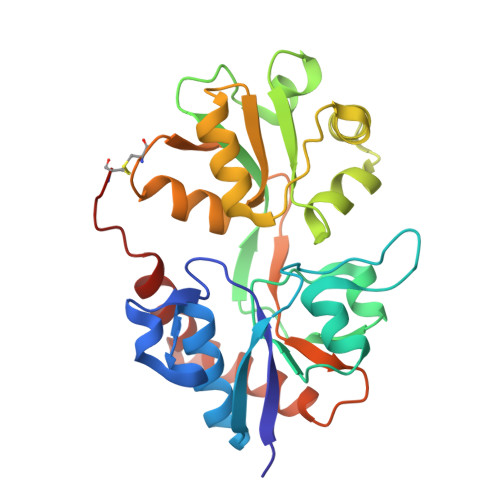
    The alpha-amino-3-hydroxy-5-methyl-4-isoxazolepropionic acid (AMPA) class of ionotropic glutamate receptors comprises four different subunits: iGluR1/iGluR2 and iGluR3/iGluR4 forming two subgroups. Three-dimensional structures have been reported only of the ligand-binding core of iGluR2. Here, we present two X-ray structures of a soluble construct of the R/G unedited flip splice variant of the ligand-binding core of iGluR4 (iGluR4(i)(R)-S1S2) in complex with glutamate or AMPA. Subtle, but important differences are found in the ligand-binding cavity between the two AMPA receptor subgroups at position 724 (Tyr in iGluR1/iGluR2 and Phe in iGluR3/iGluR4), which in iGluR4 may lead to displacement of a water molecule and hence points to the possibility to make subgroup specific ligands.


  • Organizational Affiliation

    Department of Medicinal Chemistry, Faculty of Pharmaceutical Sciences, University of Copenhagen, Universitetsparken 2, DK-2100 Copenhagen, Denmark.


Macromolecules
Find similar proteins by:  (by identity cutoff)  |  3D Structure
Entity ID: 1
MoleculeChains Sequence LengthOrganismDetailsImage
Glutamate receptor 4
A, B, C
260Rattus norvegicusMutation(s): 0 
UniProt
Find proteins for P19493 (Rattus norvegicus)
Explore P19493 
Go to UniProtKB:  P19493
Entity Groups  
Sequence Clusters30% Identity50% Identity70% Identity90% Identity95% Identity100% Identity
UniProt GroupP19493
Sequence Annotations
Expand
  • Reference Sequence
Small Molecules
Ligands 4 Unique
IDChains Name / Formula / InChI Key2D Diagram3D Interactions
AMQ
Query on AMQ

Download Ideal Coordinates CCD File 
D [auth A],
K [auth B],
T [auth C]
(S)-ALPHA-AMINO-3-HYDROXY-5-METHYL-4-ISOXAZOLEPROPIONIC ACID
C7 H10 N2 O4
UUDAMDVQRQNNHZ-YFKPBYRVSA-N
SO4
Query on SO4

Download Ideal Coordinates CCD File 
E [auth A]
F [auth A]
G [auth A]
H [auth A]
L [auth B]
E [auth A],
F [auth A],
G [auth A],
H [auth A],
L [auth B],
U [auth C],
V [auth C]
SULFATE ION
O4 S
QAOWNCQODCNURD-UHFFFAOYSA-L
GOL
Query on GOL

Download Ideal Coordinates CCD File 
I [auth A]
M [auth B]
N [auth B]
O [auth B]
P [auth B]
I [auth A],
M [auth B],
N [auth B],
O [auth B],
P [auth B],
W [auth C],
X [auth C],
Y [auth C],
Z [auth C]
GLYCEROL
C3 H8 O3
PEDCQBHIVMGVHV-UHFFFAOYSA-N
ACY
Query on ACY

Download Ideal Coordinates CCD File 
AA [auth C]
BA [auth C]
J [auth A]
Q [auth B]
R [auth B]
AA [auth C],
BA [auth C],
J [auth A],
Q [auth B],
R [auth B],
S [auth B]
ACETIC ACID
C2 H4 O2
QTBSBXVTEAMEQO-UHFFFAOYSA-N
Binding Affinity Annotations 
IDSourceBinding Affinity
AMQ Binding MOAD:  3FAT Kd: 22.8 (nM) from 1 assay(s)
PDBBind:  3FAT Kd: 22.8 (nM) from 1 assay(s)
Experimental Data & Validation

Experimental Data

  • Method: X-RAY DIFFRACTION
  • Resolution: 1.90 Å
  • R-Value Free: 0.216 
  • R-Value Work: 0.175 
  • R-Value Observed: 0.177 
  • Space Group: C 1 2 1
Unit Cell:
Length ( Å )Angle ( ˚ )
a = 105.175α = 90
b = 169.531β = 120.58
c = 73.883γ = 90
Software Package:
Software NamePurpose
SCALAdata scaling
PHASERphasing
PHENIXrefinement
PDB_EXTRACTdata extraction
MAR345dtbdata collection
MOSFLMdata reduction

Structure Validation

View Full Validation Report


Ligand Structure Quality Assessment 


Entry History 

Deposition Data

Revision History  (Full details and data files)

  • Version 1.0: 2008-12-09
    Type: Initial release
  • Version 1.1: 2011-07-13
    Changes: Non-polymer description, Version format compliance
  • Version 1.2: 2017-08-09
    Changes: Data collection, Refinement description, Source and taxonomy
  • Version 1.3: 2023-11-01
    Changes: Data collection, Database references, Derived calculations, Refinement description