3F8K

Crystal structure of protein acetyltransferase (PAT) from Sulfolobus solfataricus


Experimental Data Snapshot

  • Method: X-RAY DIFFRACTION
  • Resolution: 1.84 Å
  • R-Value Free: 0.221 
  • R-Value Work: 0.202 
  • R-Value Observed: 0.202 

wwPDB Validation   3D Report Full Report


Ligand Structure Quality Assessment 


This is version 1.3 of the entry. See complete history


Literature

Structure and Biochemical Characterization of Protein Acetyltransferase from Sulfolobus solfataricus.

Brent, M.M.Iwata, A.Carten, J.Zhao, K.Marmorstein, R.

(2009) J Biol Chem 284: 19412-19419

  • DOI: https://doi.org/10.1074/jbc.M109.014951
  • Primary Citation of Related Structures:  
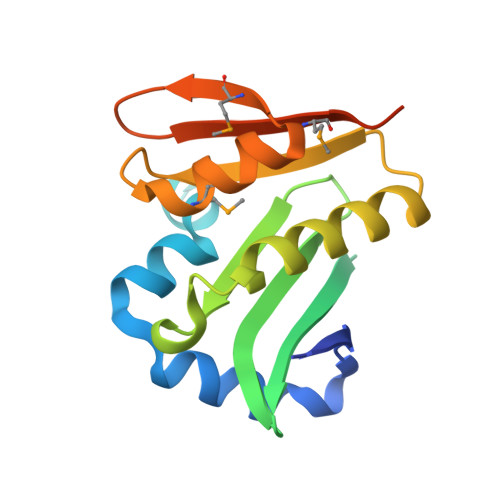
    3F8K

  • PubMed Abstract: 

    The Sulfolobus solfataricus protein acetyltransferase (PAT) acetylates ALBA, an abundant nonspecific DNA-binding protein, on Lys(16) to reduce its DNA affinity, and the Sir2 deacetylase reverses the modification to cause transcriptional repression. This represents a "primitive" model for chromatin regulation analogous to histone modification in eukaryotes. We report the 1.84-A crystal structure of PAT in complex with coenzyme A. The structure reveals homology to both prokaryotic GNAT acetyltransferases and eukaryotic histone acetyltransferases (HATs), with an additional "bent helix" proximal to the substrate binding site that might play an autoregulatory function. Investigation of active site mutants suggests that PAT does not use a single general base or acid residue for substrate deprotonation and product reprotonation, respectively, and that a diffusional step, such as substrate binding, may be rate-limiting. The catalytic efficiency of PAT toward ALBA is low relative to other acetyltransferases, suggesting that there may be better, unidentified substrates for PAT. The structural similarity of PAT to eukaryotic HATs combined with its conserved role in chromatin regulation suggests that PAT is evolutionarily related to the eukaryotic HATs.


  • Organizational Affiliation

    Wistar Institute, University of Pennsylvania, Philadelphia, Pennsylvania 19104, USA.


Macromolecules
Find similar proteins by:  (by identity cutoff)  |  3D Structure
Entity ID: 1
MoleculeChains Sequence LengthOrganismDetailsImage
Protein acetyltransferase160Saccharolobus solfataricus P2Mutation(s): 0 
Gene Names: SSO2813
UniProt
Find proteins for Q97V23 (Saccharolobus solfataricus (strain ATCC 35092 / DSM 1617 / JCM 11322 / P2))
Explore Q97V23 
Go to UniProtKB:  Q97V23
Entity Groups  
Sequence Clusters30% Identity50% Identity70% Identity90% Identity95% Identity100% Identity
UniProt GroupQ97V23
Sequence Annotations
Expand
  • Reference Sequence
Small Molecules
Ligands 1 Unique
IDChains Name / Formula / InChI Key2D Diagram3D Interactions
COA
Query on COA

Download Ideal Coordinates CCD File 
B [auth A]COENZYME A
C21 H36 N7 O16 P3 S
RGJOEKWQDUBAIZ-IBOSZNHHSA-N
Modified Residues  1 Unique
IDChains TypeFormula2D DiagramParent
MSE
Query on MSE
A
L-PEPTIDE LINKINGC5 H11 N O2 SeMET
Experimental Data & Validation

Experimental Data

  • Method: X-RAY DIFFRACTION
  • Resolution: 1.84 Å
  • R-Value Free: 0.221 
  • R-Value Work: 0.202 
  • R-Value Observed: 0.202 
  • Space Group: P 21 21 21
Unit Cell:
Length ( Å )Angle ( ˚ )
a = 44.701α = 90
b = 46.75β = 90
c = 68.624γ = 90
Software Package:
Software NamePurpose
MAR345data collection
SOLVEphasing
CNSrefinement
HKL-2000data reduction
HKL-2000data scaling

Structure Validation

View Full Validation Report


Ligand Structure Quality Assessment 


Entry History 

Deposition Data

  • Released Date: 2009-05-26 
  • Deposition Author(s): Brent, M.M.

Revision History  (Full details and data files)

  • Version 1.0: 2009-05-26
    Type: Initial release
  • Version 1.1: 2011-07-13
    Changes: Version format compliance
  • Version 1.2: 2017-11-01
    Changes: Refinement description
  • Version 1.3: 2023-12-27
    Changes: Data collection, Database references, Derived calculations