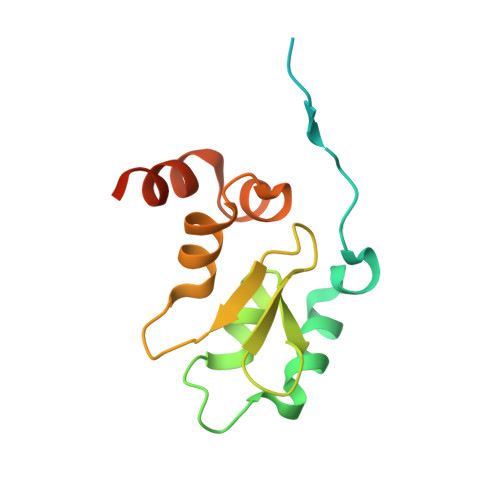
3F7G

Structure of the BIR domain from ML-IAP bound to a peptidomimetic


Experimental Data Snapshot

  • Method: X-RAY DIFFRACTION
  • Resolution: 2.30 Å
  • R-Value Free: 0.209 
  • R-Value Work: 0.167 
  • R-Value Observed: 0.169 

wwPDB Validation   3D Report Full Report


Ligand Structure Quality Assessment 


This is version 1.3 of the entry. See complete history


Literature

Orally bioavailable antagonists of inhibitor of apoptosis proteins based on an azabicyclooctane scaffold

Cohen, F.Alicke, B.Elliott, L.O.Flygare, J.A.Goncharov, T.Keteltas, S.F.Franklin, M.C.Frankovitz, S.Stephan, J.P.Tsui, V.Vucic, D.Wong, H.Fairbrother, W.J.

(2009) J Med Chem 52: 1723-1730

  • DOI: https://doi.org/10.1021/jm801450c
  • Primary Citation of Related Structures:  
    3F7G, 3F7H, 3F7I

  • PubMed Abstract: 

    A series of IAP antagonists based on an azabicyclooctane scaffold was designed and synthesized. The most potent of these compounds, 14b, binds to the XIAP BIR3 domain, the BIR domain of ML-IAP, and the BIR3 domain of c-IAP1 with K(i) values of 140, 38, and 33 nM, respectively. These compounds promote degradation of c-IAP1, activate caspases, and lead to decreased viability of breast cancer cells without affecting normal mammary epithelial cells. Finally, compound 14b inhibits tumor growth when dosed orally in a breast cancer xenograft model.


  • Organizational Affiliation

    Departments of Discovery Chemistry, Genentech, Inc., 1 DNA Way, South San Francisco, California 94080, USA. fcohen@gene.com


Macromolecules
Find similar proteins by:  (by identity cutoff)  |  3D Structure
Entity ID: 1
MoleculeChains Sequence LengthOrganismDetailsImage
Baculoviral IAP repeat-containing protein 7
A, B, C, D, E
140Homo sapiensMutation(s): 0 
Gene Names: also known as KIAP OR MLIAP OR LIVINBIRC7KIAPLIVINMLIAPRNF50UNQ5800/PRO19607/PRO21344
UniProt & NIH Common Fund Data Resources
Find proteins for Q96CA5 (Homo sapiens)
Explore Q96CA5 
Go to UniProtKB:  Q96CA5
PHAROS:  Q96CA5
GTEx:  ENSG00000101197 
Entity Groups  
Sequence Clusters30% Identity50% Identity70% Identity90% Identity95% Identity100% Identity
UniProt GroupQ96CA5
Sequence Annotations
Expand
  • Reference Sequence
Small Molecules
Binding Affinity Annotations 
IDSourceBinding Affinity
389 BindingDB:  3F7G Ki: 43 (nM) from 1 assay(s)
PDBBind:  3F7G Ki: 43 (nM) from 1 assay(s)
Experimental Data & Validation

Experimental Data

  • Method: X-RAY DIFFRACTION
  • Resolution: 2.30 Å
  • R-Value Free: 0.209 
  • R-Value Work: 0.167 
  • R-Value Observed: 0.169 
  • Space Group: P 32
Unit Cell:
Length ( Å )Angle ( ˚ )
a = 84.016α = 90
b = 84.016β = 90
c = 94.476γ = 120
Software Package:
Software NamePurpose
HKL-2000data collection
REFMACrefinement
HKL-2000data reduction
HKL-2000data scaling

Structure Validation

View Full Validation Report


Ligand Structure Quality Assessment 


Entry History 

Deposition Data

Revision History  (Full details and data files)

  • Version 1.0: 2009-03-17
    Type: Initial release
  • Version 1.1: 2011-07-13
    Changes: Advisory, Refinement description, Version format compliance
  • Version 1.2: 2017-10-25
    Changes: Refinement description
  • Version 1.3: 2023-09-06
    Changes: Data collection, Database references, Derived calculations, Refinement description, Structure summary