3F7D

SF-1 LBD bound by phosphatidylcholine


Experimental Data Snapshot

  • Method: X-RAY DIFFRACTION
  • Resolution: 2.20 Å
  • R-Value Free: 0.241 
  • R-Value Work: 0.180 
  • R-Value Observed: 0.182 

wwPDB Validation   3D Report Full Report


Ligand Structure Quality Assessment 


This is version 1.5 of the entry. See complete history


Literature

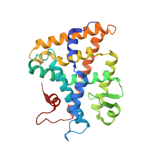
Structure of SF-1 bound by different phospholipids: evidence for regulatory ligands.

Sablin, E.P.Blind, R.D.Krylova, I.N.Ingraham, J.G.Cai, F.Williams, J.D.Fletterick, R.J.Ingraham, H.A.

(2009) Mol Endocrinol 23: 25-34

  • DOI: https://doi.org/10.1210/me.2007-0508
  • Primary Citation of Related Structures:  
    3F7D

  • PubMed Abstract: 

    Despite the fact that many nuclear receptors are ligand dependent, the existence of obligate regulatory ligands is debated for some receptors, including steroidogenic factor 1 (SF-1). Although fortuitously bound bacterial phospholipids were discovered in the structures of the SF-1 ligand-binding domain (LBD), these lipids might serve merely as structural ligands. Thus, we examined whether exogenously added phospholipids would exchange for these bacterial lipids and bind to SF-1. Here, we report the first crystal structure of the SF-1 LBD bound by the exchanged phosphatidylcholine. Although the bound phosphatidylcholine phospholipid mimics the conformation of bound bacterial phosphoplipids, two surface loops, L2-3 and L11-12, surrounding the entrance to the pocket vary significantly between different SF-1 LBD structures. Based on this observation, we hypothesized that a bound ligand might control the conformations of loops L2-3 and L11-12, and that conserved residues in these dynamic loops could influence ligand binding and the receptor function. Consistent with this hypothesis, impaired phospholipid exchange and diminished transcriptional activity were observed for loop L11-12 SF-1 mutants and for the loop L2-3 human mutant R255L. The endocrine disease associated with this L2-3 mutation coupled with our cellular and biochemical data suggest that critical residues at the mouth of the ligand-binding pocket have evolved for efficient binding of phospholipid ligands and for achieving optimal SF-1 activity.


  • Organizational Affiliation

    Department of Biochemistry and Biophysics, University of California, San Francisco, California 94143, USA.


Macromolecules
Find similar proteins by:  (by identity cutoff)  |  3D Structure
Entity ID: 1
MoleculeChains Sequence LengthOrganismDetailsImage
nuclear receptor SF-1244Mus musculusMutation(s): 4 
Gene Names: Nr5a1Ftzf1RP23-354G20.5-001
UniProt
Find proteins for P33242 (Mus musculus)
Explore P33242 
Go to UniProtKB:  P33242
Entity Groups  
Sequence Clusters30% Identity50% Identity70% Identity90% Identity95% Identity100% Identity
UniProt GroupP33242
Sequence Annotations
Expand
  • Reference Sequence

Find similar proteins by:  Sequence   |   3D Structure  

Entity ID: 2
MoleculeChains Sequence LengthOrganismDetailsImage
Peroxisome proliferator-activated receptor gamma coactivator 1-alpha14Mus musculusMutation(s): 0 
UniProt & NIH Common Fund Data Resources
Find proteins for O70343 (Mus musculus)
Explore O70343 
Go to UniProtKB:  O70343
IMPC:  MGI:1342774
Entity Groups  
Sequence Clusters30% Identity50% Identity70% Identity90% Identity95% Identity100% Identity
UniProt GroupO70343
Sequence Annotations
Expand
  • Reference Sequence
Small Molecules
Ligands 1 Unique
IDChains Name / Formula / InChI Key2D Diagram3D Interactions
P42
Query on P42

Download Ideal Coordinates CCD File 
C [auth A](2S)-2-{[(1R)-1-hydroxyhexadecyl]oxy}-3-{[(1R)-1-hydroxyoctadecyl]oxy}propyl 2-(trimethylammonio)ethyl phosphate
C42 H88 N O8 P
LYKCEYRWVRQPBD-FEWNNGCESA-N
Modified Residues  1 Unique
IDChains TypeFormula2D DiagramParent
CAF
Query on CAF
A
L-PEPTIDE LINKINGC5 H12 As N O3 SCYS
Experimental Data & Validation

Experimental Data

  • Method: X-RAY DIFFRACTION
  • Resolution: 2.20 Å
  • R-Value Free: 0.241 
  • R-Value Work: 0.180 
  • R-Value Observed: 0.182 
  • Space Group: P 21 21 21
Unit Cell:
Length ( Å )Angle ( ˚ )
a = 46.895α = 90
b = 67.228β = 90
c = 82.689γ = 90
Software Package:
Software NamePurpose
PHENIXrefinement
CNSrefinement
ADSCdata collection
DENZOdata reduction
SCALEPACKdata scaling
CNSphasing

Structure Validation

View Full Validation Report


Ligand Structure Quality Assessment 


Entry History 

Deposition Data

Revision History  (Full details and data files)

  • Version 1.0: 2008-12-09
    Type: Initial release
  • Version 1.1: 2011-07-13
    Changes: Version format compliance
  • Version 1.2: 2014-04-02
    Changes: Source and taxonomy
  • Version 1.3: 2019-07-24
    Changes: Data collection, Derived calculations, Refinement description
  • Version 1.4: 2021-10-20
    Changes: Database references, Derived calculations
  • Version 1.5: 2023-09-06
    Changes: Data collection, Refinement description