Conformational Closure of the Catalytic Site of Human CD38 Induced by Calcium
Liu, Q., Graeff, R., Kriksunov, I.A., Lam, C.M.C., Lee, H.C., Hao, Q.(2008) Biochemistry 47: 13966-13973
- PubMed: 19117080
- Primary Citation of Related Structures:
3F6Y - PubMed Abstract:
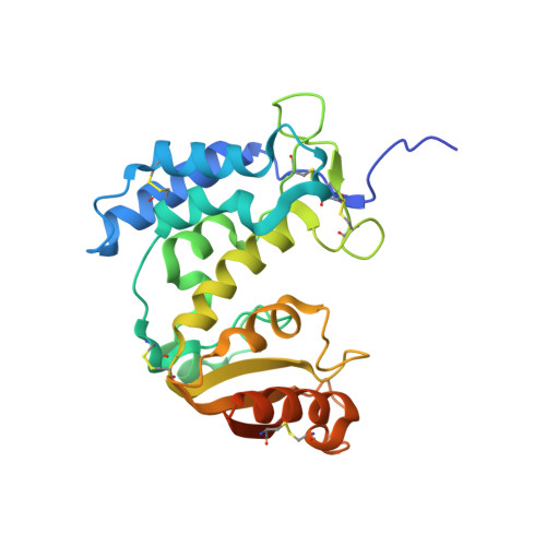
First identified on the surface of lymphoids as a type II transmembrane protein, CD38 has now been established to have dual functions not only as a receptor but also as a multifunctional enzyme,catalyzing the synthesis of and hydrolysis of a general calcium messenger molecule, cyclic ADP-ribose(cADPR). The receptorial functions of CD38 include the induction of cell adhesion, differentiation,apoptosis, and cytokine production upon antibody ligation. Here we determined the crystal structure of calcium-loaded human CD38 at 1.45 A resolution which reveals that CD38 undergoes dramatic structural changes to an inhibited conformation in the presence of calcium. The structural changes are highly localized and occur in only two regions. The first region is part of the active site and consists of residues 121-141.In the presence of calcium, W125 moves 5 A into the active site and forms hydrophobic interactions with W189. The movement closes the active site pocket and reduces entry of substrates, resulting in inhibition of the enzymatic activity. The structural role of calcium in inducing these conformational changes is readily visualized in the crystal structure. The other region that undergoes calcium-induced changes is at the receptor region, where a highly ordered helix is unraveled to a random coil. The results suggest a novel conformational coupling mechanism, whereby protein interaction targeted at the receptor region can effectively regulate the enzymatic activity of CD38.
Organizational Affiliation:
MacCHESS, Cornell High Energy Synchrotron Source, and School of Applied and Engineering Physics, Cornell University, Ithaca, New York 14853, USA.