3F5M

Crystal Structure of ATP-Bound Phosphofructokinase from Trypanosoma brucei


Experimental Data Snapshot

  • Method: X-RAY DIFFRACTION
  • Resolution: 2.70 Å
  • R-Value Free: 0.286 
  • R-Value Work: 0.224 
  • R-Value Observed: 0.227 

wwPDB Validation   3D Report Full Report


Ligand Structure Quality Assessment 


This is version 1.2 of the entry. See complete history


Literature

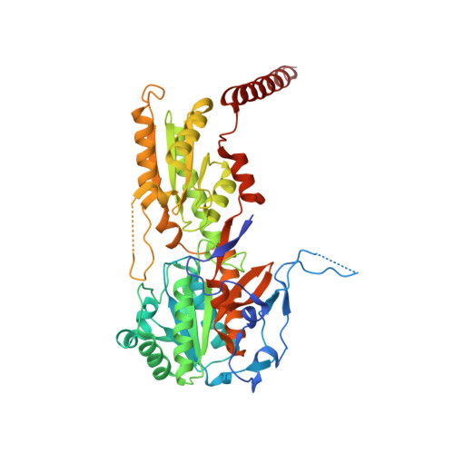
The crystal structure of ATP-bound phosphofructokinase from Trypanosoma brucei reveals conformational transitions different from those of other phosphofructokinases.

McNae, I.W.Martinez-Oyanedel, J.Keillor, J.W.Michels, P.A.Fothergill-Gilmore, L.A.Walkinshaw, M.D.

(2009) J Mol Biol 385: 1519-1533

  • DOI: https://doi.org/10.1016/j.jmb.2008.11.047
  • Primary Citation of Related Structures:  
    3F5M

  • PubMed Abstract: 

    The crystal structure of the ATP-bound form of the tetrameric phosphofructokinase (PFK) from Trypanosoma brucei enables detailed comparisons to be made with the structures of the apoenzyme form of the same enzyme, as well as with those of bacterial ATP-dependent and PP(i)-dependent PFKs. The active site of T. brucei PFK (which is strictly ATP-dependent but belongs to the PP(i)-dependent family by sequence similarities) is a chimera of the two types of PFK. In particular, the active site of T. brucei PFK possesses amino acid residues and structural features characteristic of both types of PFK. Conformational changes upon ATP binding are observed that include the opening of the active site to accommodate the two substrates, MgATP and fructose 6-phosphate, and a dramatic ordering of the C-terminal helices, which act like reaching arms to hold the tetramer together. These conformational transitions are fundamentally different from those of other ATP-dependent PFKs. The substantial differences in structure and mechanism of T. brucei PFK compared with bacterial and mammalian PFKs give optimism for the discovery of species-specific drugs for the treatment of diseases caused by protist parasites of the trypanosomatid family.


  • Organizational Affiliation

    Structural Biochemistry Group, Institute of Structural and Molecular Biology, University of Edinburgh, King's Buildings, Edinburgh, Scotland.


Macromolecules
Find similar proteins by:  (by identity cutoff)  |  3D Structure
Entity ID: 1
MoleculeChains Sequence LengthOrganismDetailsImage
6-phospho-1-fructokinase (ATP-dependent phosphofructokinase)
A, B, C, D
487Trypanosoma bruceiMutation(s): 0 
EC: 2.7.1.11
UniProt
Find proteins for O15648 (Trypanosoma brucei brucei)
Explore O15648 
Go to UniProtKB:  O15648
Entity Groups  
Sequence Clusters30% Identity50% Identity70% Identity90% Identity95% Identity100% Identity
UniProt GroupO15648
Sequence Annotations
Expand
  • Reference Sequence
Small Molecules
Ligands 5 Unique
IDChains Name / Formula / InChI Key2D Diagram3D Interactions
ATP
Query on ATP

Download Ideal Coordinates CCD File 
E [auth A],
J [auth B],
M [auth C],
Q [auth D]
ADENOSINE-5'-TRIPHOSPHATE
C10 H16 N5 O13 P3
ZKHQWZAMYRWXGA-KQYNXXCUSA-N
SO4
Query on SO4

Download Ideal Coordinates CCD File 
O [auth C]SULFATE ION
O4 S
QAOWNCQODCNURD-UHFFFAOYSA-L
GOL
Query on GOL

Download Ideal Coordinates CCD File 
G [auth A],
N [auth C]
GLYCEROL
C3 H8 O3
PEDCQBHIVMGVHV-UHFFFAOYSA-N
MG
Query on MG

Download Ideal Coordinates CCD File 
F [auth A],
K [auth B],
R [auth D]
MAGNESIUM ION
Mg
JLVVSXFLKOJNIY-UHFFFAOYSA-N
NA
Query on NA

Download Ideal Coordinates CCD File 
H [auth A],
I [auth A],
L [auth B],
P [auth C]
SODIUM ION
Na
FKNQFGJONOIPTF-UHFFFAOYSA-N
Experimental Data & Validation

Experimental Data

  • Method: X-RAY DIFFRACTION
  • Resolution: 2.70 Å
  • R-Value Free: 0.286 
  • R-Value Work: 0.224 
  • R-Value Observed: 0.227 
  • Space Group: P 21 21 21
Unit Cell:
Length ( Å )Angle ( ˚ )
a = 96.578α = 90
b = 117.57β = 90
c = 176.587γ = 90
Software Package:
Software NamePurpose
PHENIXrefinement

Structure Validation

View Full Validation Report


Ligand Structure Quality Assessment 


Entry History 

Deposition Data

Revision History  (Full details and data files)

  • Version 1.0: 2008-11-25
    Type: Initial release
  • Version 1.1: 2011-07-13
    Changes: Non-polymer description, Version format compliance
  • Version 1.2: 2023-11-01
    Changes: Data collection, Database references, Derived calculations, Refinement description