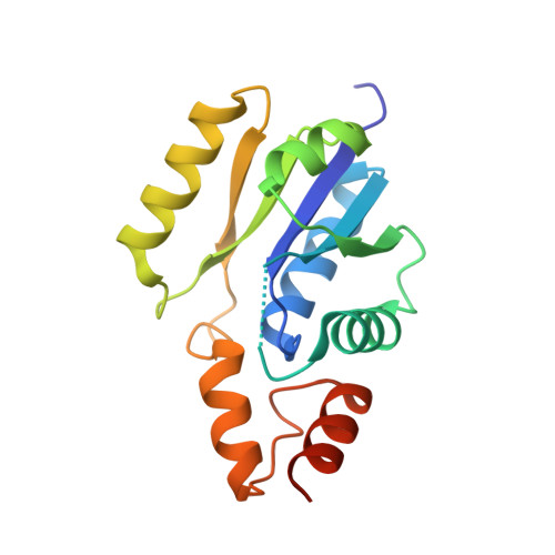
3F3M

Six Crystal Structures of Two Phosphopantetheine Adenylyltransferases Reveal an Alternative Ligand Binding Mode and an Associated Structural Change


Experimental Data Snapshot

  • Method: X-RAY DIFFRACTION
  • Resolution: 2.40 Å
  • R-Value Free: 0.250 
  • R-Value Work: 0.211 

wwPDB Validation   3D Report Full Report


This is version 1.3 of the entry. See complete history


Literature

The structure of Staphylococcus aureus phosphopantetheine adenylyltransferase in complex with 3'-phosphoadenosine 5'-phosphosulfate reveals a new ligand-binding mode

Lee, H.H.Yoon, H.J.Kang, J.Y.Park, J.H.Kim, D.J.Choi, K.H.Lee, S.K.Song, J.Kim, H.J.Suh, S.W.

(2009) Acta Crystallogr Sect F Struct Biol Cryst Commun 65: 987-991

  • DOI: https://doi.org/10.1107/S1744309109036616
  • Primary Citation of Related Structures:  
    3F3M

  • PubMed Abstract: 

    Bacterial phosphopantetheine adenylyltransferase (PPAT) catalyzes the penultimate step in the coenzyme A (CoA) biosynthetic pathway. It catalyzes the reversible transfer of an adenylyl group from ATP to 4'-phosphopantetheine (Ppant) to form dephospho-CoA (dPCoA) and pyrophosphate. Previous structural studies have revealed how several ligands are recognized by bacterial PPATs. ATP, ADP, Ppant and dPCoA bind to the same binding site in a highly similar manner, while CoA binds to a partially overlapping site in a different mode. To provide further structural insights into ligand binding, the crystal structure of Staphylococcus aureus PPAT was solved in a binary complex with 3'-phosphoadenosine 5'-phosphosulfate (PAPS). This study unexpectedly revealed a new mode of ligand binding to PPAT, thus providing potentially useful information for structure-based discovery of inhibitors of bacterial PPATs.


  • Organizational Affiliation

    Department of Chemistry, College of Natural Sciences, Seoul National University, Seoul 151-742, Republic of Korea.


Macromolecules
Find similar proteins by:  (by identity cutoff)  |  3D Structure
Entity ID: 1
MoleculeChains Sequence LengthOrganismDetailsImage
Phosphopantetheine adenylyltransferase168Staphylococcus aureusMutation(s): 0 
Gene Names: coaD
EC: 2.7.7.3
UniProt
Find proteins for P63820 (Staphylococcus aureus (strain MW2))
Explore P63820 
Go to UniProtKB:  P63820
Entity Groups  
Sequence Clusters30% Identity50% Identity70% Identity90% Identity95% Identity100% Identity
UniProt GroupP63820
Sequence Annotations
Expand
  • Reference Sequence
Small Molecules
Ligands 1 Unique
IDChains Name / Formula / InChI Key2D Diagram3D Interactions
PPS
Query on PPS

Download Ideal Coordinates CCD File 
B [auth A]3'-PHOSPHATE-ADENOSINE-5'-PHOSPHATE SULFATE
C10 H15 N5 O13 P2 S
GACDQMDRPRGCTN-KQYNXXCUSA-N
Experimental Data & Validation

Experimental Data

  • Method: X-RAY DIFFRACTION
  • Resolution: 2.40 Å
  • R-Value Free: 0.250 
  • R-Value Work: 0.211 
  • Space Group: P 63 2 2
Unit Cell:
Length ( Å )Angle ( ˚ )
a = 66.774α = 90
b = 66.774β = 90
c = 124.134γ = 120
Software Package:
Software NamePurpose
CNSrefinement
PDB_EXTRACTdata extraction
HKL-2000data collection
HKL-2000data reduction
HKL-2000data scaling
CNSphasing

Structure Validation

View Full Validation Report


Entry History 

Deposition Data

Revision History  (Full details and data files)

  • Version 1.0: 2009-10-20
    Type: Initial release
  • Version 1.1: 2011-07-13
    Changes: Version format compliance
  • Version 1.2: 2012-04-25
    Changes: Database references
  • Version 1.3: 2023-11-01
    Changes: Data collection, Database references, Derived calculations, Refinement description