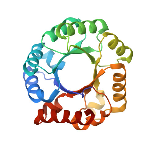
3EXR

Crystal structure of KGPDC from Streptococcus mutans


Experimental Data Snapshot

  • Method: X-RAY DIFFRACTION
  • Resolution: 1.70 Å
  • R-Value Free: 0.244 
  • R-Value Work: 0.206 
  • R-Value Observed: 0.208 

wwPDB Validation   3D Report Full Report


This is version 1.3 of the entry. See complete history


Literature

Open-closed conformational change revealed by the crystal structures of 3-keto-L-gulonate 6-phosphate decarboxylase from Streptococcus mutans

Li, G.L.Liu, X.Nan, J.Brostromer, E.Li, L.F.Su, X.D.

(2009) Biochem Biophys Res Commun 381: 429-433

  • DOI: https://doi.org/10.1016/j.bbrc.2009.02.049
  • Primary Citation of Related Structures:  
    3EXR, 3EXS, 3EXT

  • PubMed Abstract: 

    The 3-keto-L-gulonate 6-phosphate decarboxylase (KGPDC) catalyses the decarboxylation of 3-keto-L-gulonate 6-phosphate to L-xylulose in the presence of magnesium ions. The enzyme is involved in L-ascorbate metabolism and plays an essential role in the pathway of glucuronate interconversion. Crystal structures of Streptococcus mutans KGPDC were determined in the absence and presence of the product analog D-ribulose 5-phosphate. We have observed an 8 A alphaB-helix movement and other structural rearrangements around the active site between the apo-structures and product analog bound structure. These drastic conformational changes upon ligand binding are the first observation of this kind for the KGPDC family. The flexibilities of both the alpha-helix lid and the side chains of Arg144 and Arg197 are associated with substrate binding and product releasing. The open-closed conformational changes of the active site, through the movements of the alpha-helix lid and the arginine residues are important for substrate binding and catalysis.


  • Organizational Affiliation

    The National Laboratory of Protein Engineering and Plant Genetic Engineering, College of Life Science, Peking University, Beijing 100871, People's Republic of China.


Macromolecules
Find similar proteins by:  (by identity cutoff)  |  3D Structure
Entity ID: 1
MoleculeChains Sequence LengthOrganismDetailsImage
RmpD (Hexulose-6-phosphate synthase)
A, B, C, D
221Streptococcus mutansMutation(s): 0 
Gene Names: ulaD
EC: 4.1.1.85
UniProt
Find proteins for Q93DA8 (Streptococcus mutans)
Explore Q93DA8 
Go to UniProtKB:  Q93DA8
Entity Groups  
Sequence Clusters30% Identity50% Identity70% Identity90% Identity95% Identity100% Identity
UniProt GroupQ93DA8
Sequence Annotations
Expand
  • Reference Sequence
Experimental Data & Validation

Experimental Data

  • Method: X-RAY DIFFRACTION
  • Resolution: 1.70 Å
  • R-Value Free: 0.244 
  • R-Value Work: 0.206 
  • R-Value Observed: 0.208 
  • Space Group: P 21 21 21
Unit Cell:
Length ( Å )Angle ( ˚ )
a = 59.83α = 90
b = 127.23β = 90
c = 130.77γ = 90
Software Package:
Software NamePurpose
XSCALEdata scaling
REFMACrefinement
PDB_EXTRACTdata extraction

Structure Validation

View Full Validation Report


Entry History 

Deposition Data

Revision History  (Full details and data files)

  • Version 1.0: 2009-08-25
    Type: Initial release
  • Version 1.1: 2011-07-13
    Changes: Version format compliance
  • Version 1.2: 2018-05-23
    Changes: Data collection, Refinement description
  • Version 1.3: 2023-11-01
    Changes: Data collection, Database references, Refinement description