


The crystal structure of the DNA binding protein from Silicibacter pomeroyi
Zhang, R., Li, H., Freeman, L., Joachimiak, A.To be published.
Experimental Data Snapshot
wwPDB Validation 3D Report Full Report

Entity ID: 1 | |||||
|---|---|---|---|---|---|
| Molecule | Chains | Sequence Length | Organism | Details | Image |
| DNA-binding protein | 86 | Ruegeria pomeroyi | Mutation(s): 0 Gene Names: GI:56696110, SPO1217 | 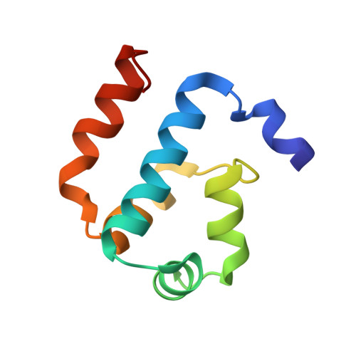 | |
UniProt | |||||
Find proteins for Q5LU41 (Ruegeria pomeroyi (strain ATCC 700808 / DSM 15171 / DSS-3)) Explore Q5LU41 Go to UniProtKB: Q5LU41 | |||||
Entity Groups | |||||
| Sequence Clusters | 30% Identity50% Identity70% Identity90% Identity95% Identity100% Identity | ||||
| UniProt Group | Q5LU41 | ||||
Sequence AnnotationsExpand | |||||
| |||||
| Length ( Å ) | Angle ( ˚ ) |
|---|---|
| a = 40.164 | α = 90 |
| b = 60.641 | β = 90 |
| c = 68.506 | γ = 90 |
| Software Name | Purpose |
|---|---|
| SBC-Collect | data collection |
| HKL-3000 | phasing |
| REFMAC | refinement |
| HKL-3000 | data reduction |
| HKL-3000 | data scaling |
RCSB PDB (citation) is hosted by
RCSB PDB is a member of the

