3EUH

Crystal Structure of the MukE-MukF Complex


Experimental Data Snapshot

  • Method: X-RAY DIFFRACTION
  • Resolution: 2.90 Å
  • R-Value Free: 0.274 
  • R-Value Work: 0.225 

wwPDB Validation   3D Report Full Report


This is version 1.3 of the entry. See complete history


Literature

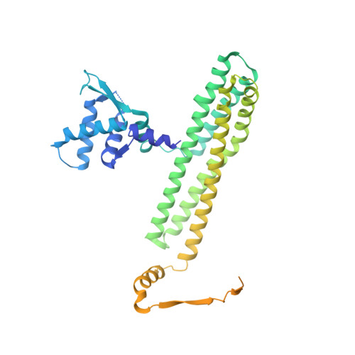
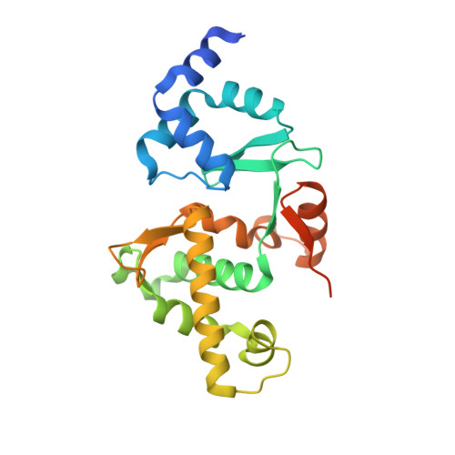
Structural studies of a bacterial condensin complex reveal ATP-dependent disruption of intersubunit interactions.

Woo, J.S.Lim, J.H.Shin, H.C.Suh, M.K.Ku, B.Lee, K.H.Joo, K.Robinson, H.Lee, J.Park, S.Y.Ha, N.C.Oh, B.H.

(2009) Cell 136: 85-96

  • DOI: https://doi.org/10.1016/j.cell.2008.10.050
  • Primary Citation of Related Structures:  
    3EUH, 3EUJ, 3EUK

  • PubMed Abstract: 

    Condensins are key mediators of chromosome condensation across organisms. Like other condensins, the bacterial MukBEF condensin complex consists of an SMC family protein dimer containing two ATPase head domains, MukB, and two interacting subunits, MukE and MukF. We report complete structural views of the intersubunit interactions of this condensin along with ensuing studies that reveal a role for the ATPase activity of MukB. MukE and MukF together form an elongated dimeric frame, and MukF's C-terminal winged-helix domains (C-WHDs) bind MukB heads to constitute closed ring-like structures. Surprisingly, one of the two bound C-WHDs is forced to detach upon ATP-mediated engagement of MukB heads. This detachment reaction depends on the linker segment preceding the C-WHD, and mutations on the linker restrict cell growth. Thus ATP-dependent transient disruption of the MukB-MukF interaction, which creates openings in condensin ring structures, is likely to be a critical feature of the functional mechanism of condensins.


  • Organizational Affiliation

    Center for Biomolecular Recognition and Division of Molecular and Life Science, Department of Life Sciences, Pohang University of Science and Technology, Pohang, Kyungbuk, 790-784, Korea.


Macromolecules
Find similar proteins by:  (by identity cutoff)  |  3D Structure
Entity ID: 1
MoleculeChains Sequence LengthOrganismDetailsImage
Chromosome partition protein mukF
A, B
440Escherichia coli K-12Mutation(s): 0 
UniProt
Find proteins for P60293 (Escherichia coli (strain K12))
Explore P60293 
Go to UniProtKB:  P60293
Entity Groups  
Sequence Clusters30% Identity50% Identity70% Identity90% Identity95% Identity100% Identity
UniProt GroupP60293
Sequence Annotations
Expand
  • Reference Sequence
Find similar proteins by:  (by identity cutoff)  |  3D Structure
Entity ID: 2
MoleculeChains Sequence LengthOrganismDetailsImage
MukE
C, D, E, F
234Escherichia coliMutation(s): 0 
UniProt
Find proteins for P22524 (Escherichia coli (strain K12))
Explore P22524 
Go to UniProtKB:  P22524
Entity Groups  
Sequence Clusters30% Identity50% Identity70% Identity90% Identity95% Identity100% Identity
UniProt GroupP22524
Sequence Annotations
Expand
  • Reference Sequence
Experimental Data & Validation

Experimental Data

  • Method: X-RAY DIFFRACTION
  • Resolution: 2.90 Å
  • R-Value Free: 0.274 
  • R-Value Work: 0.225 
  • Space Group: P 21 21 21
Unit Cell:
Length ( Å )Angle ( ˚ )
a = 77.338α = 90
b = 151.146β = 90
c = 188.072γ = 90
Software Package:
Software NamePurpose
DENZOdata reduction
SCALEPACKdata scaling
CNSrefinement
PDB_EXTRACTdata extraction
MLPHAREphasing

Structure Validation

View Full Validation Report


Entry History 

Deposition Data

Revision History  (Full details and data files)

  • Version 1.0: 2009-01-20
    Type: Initial release
  • Version 1.1: 2011-07-13
    Changes: Version format compliance
  • Version 1.2: 2017-10-25
    Changes: Refinement description
  • Version 1.3: 2023-12-27
    Changes: Data collection, Database references, Derived calculations