


Crystal structures of isocitrate lyase from Brucella melitensis
SSGCIDTo be published.
Experimental Data Snapshot
wwPDB Validation 3D Report Full Report

Entity ID: 1 | |||||
|---|---|---|---|---|---|
| Molecule | Chains | Sequence Length | Organism | Details | Image |
| Methylisocitrate lyase | 298 | Burkholderia pseudomallei 1655 | Mutation(s): 0 Gene Names: BURPS1655_C0444, BURPS1710B_2458, BURPS1710b_3237, MSRB, prpB EC: 4.1.3.30 | 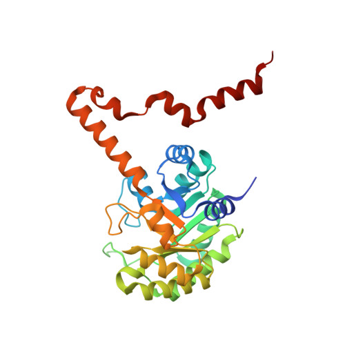 | |
Entity Groups | |||||
| Sequence Clusters | 30% Identity50% Identity70% Identity90% Identity95% Identity100% Identity | ||||
Sequence AnnotationsExpand | |||||
| |||||
| Length ( Å ) | Angle ( ˚ ) |
|---|---|
| a = 163.807 | α = 90 |
| b = 172.404 | β = 90 |
| c = 179.277 | γ = 90 |
| Software Name | Purpose |
|---|---|
| d*TREK | data scaling |
| PHASER | phasing |
| REFMAC | refinement |
| PDB_EXTRACT | data extraction |
RCSB PDB (citation) is hosted by
RCSB PDB is a member of the

