


The structure of RssB, a ClpX adaptor protein that regulates sigma S
Levchenko, I., Grant, R.A., Sauer, R.T., Baker, T.A.To be published.
Experimental Data Snapshot
wwPDB Validation 3D Report Full Report

Entity ID: 1 | |||||
|---|---|---|---|---|---|
| Molecule | Chains | Sequence Length | Organism | Details | Image |
| Protein hnr | 130 | Escherichia coli K-12 | Mutation(s): 0 Gene Names: b1235, hnr, JW1223, rssb, ychL | 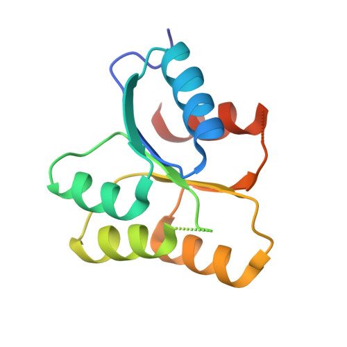 | |
UniProt | |||||
Find proteins for P0AEV1 (Escherichia coli (strain K12)) Explore P0AEV1 Go to UniProtKB: P0AEV1 | |||||
Entity Groups | |||||
| Sequence Clusters | 30% Identity50% Identity70% Identity90% Identity95% Identity100% Identity | ||||
| UniProt Group | P0AEV1 | ||||
Sequence AnnotationsExpand | |||||
| |||||
| Length ( Å ) | Angle ( ˚ ) |
|---|---|
| a = 45.114 | α = 90 |
| b = 45.114 | β = 90 |
| c = 96.797 | γ = 120 |
| Software Name | Purpose |
|---|---|
| DENZO | data reduction |
| SCALEPACK | data scaling |
| SHARP | phasing |
| DM | phasing |
| PHENIX | refinement |
| PDB_EXTRACT | data extraction |
| ADSC | data collection |
RCSB PDB (citation) is hosted by
RCSB PDB is a member of the

