3ENL

REFINED STRUCTURE OF YEAST APO-ENOLASE AT 2.25 ANGSTROMS RESOLUTION


Experimental Data Snapshot

  • Method: X-RAY DIFFRACTION
  • Resolution: 2.25 Å
  • R-Value Observed: 0.154 

wwPDB Validation   3D Report Full Report


This is version 1.4 of the entry. See complete history


Literature

Refined structure of yeast apo-enolase at 2.25 A resolution.

Stec, B.Lebioda, L.

(1990) J Mol Biol 211: 235-248

  • DOI: https://doi.org/10.1016/0022-2836(90)90023-F
  • Primary Citation of Related Structures:  
    3ENL

  • PubMed Abstract: 

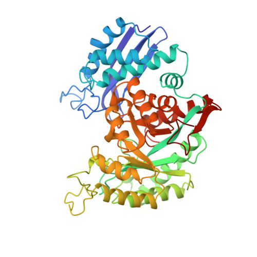
    The crystal structure of apo-enolase from baker's yeast (Saccharomyces cerevisiae) was established at 2.25 A resolution using a restrained least-squares refinement method. Based on 21,077 independent reflections of better than 8 A resolution, a final R-factor of 15.4% was obtained with a model obeying standard geometry within 0.017 A in bond length and 3.5 degrees in bond angles. The upper limit for the co-ordinate accuracy of the atoms was estimated to be 0.18 A. The refinement confirmed the heterodox, non-parallel character of the 8-fold beta alpha-barrel domain with beta beta alpha alpha(beta alpha)6 topology. The reported structure for which the data were collected at pH 5.0 represents an apo-form of the enzyme. Of the three carboxylic ligands that form the conformational metal ion binding site two, Glu295 and Asp320, are very close and presumably form a strong acidic type hydrogen bond with the proton partially replacing the electric charge of the physiological cofactor Mg2+. The single sulfate ion found in the structure is in the active site cavity, co-ordinated to the side-chains of Lys345 and Arg374, and to the N atom of Ser375. It is located about 7.4 A from the conformational metal ion binding site. It occupies the site in which the phosphate group of the substrate binds.


  • Organizational Affiliation

    Department of Chemistry, University of South Carolina, Columbia 29208.


Macromolecules
Find similar proteins by:  (by identity cutoff)  |  3D Structure
Entity ID: 1
MoleculeChains Sequence LengthOrganismDetailsImage
ENOLASE436Saccharomyces cerevisiaeMutation(s): 0 
EC: 4.2.1.11
UniProt
Find proteins for P00924 (Saccharomyces cerevisiae (strain ATCC 204508 / S288c))
Explore P00924 
Go to UniProtKB:  P00924
Entity Groups  
Sequence Clusters30% Identity50% Identity70% Identity90% Identity95% Identity100% Identity
UniProt GroupP00924
Sequence Annotations
Expand
  • Reference Sequence
Small Molecules
Ligands 1 Unique
IDChains Name / Formula / InChI Key2D Diagram3D Interactions
SO4
Query on SO4

Download Ideal Coordinates CCD File 
B [auth A]SULFATE ION
O4 S
QAOWNCQODCNURD-UHFFFAOYSA-L
Experimental Data & Validation

Experimental Data

  • Method: X-RAY DIFFRACTION
  • Resolution: 2.25 Å
  • R-Value Observed: 0.154 
  • Space Group: P 42 21 2
Unit Cell:
Length ( Å )Angle ( ˚ )
a = 124.1α = 90
b = 124.1β = 90
c = 66.9γ = 90
Software Package:
Software NamePurpose
PROLSQrefinement

Structure Validation

View Full Validation Report


Entry History 

Deposition Data

Revision History  (Full details and data files)

  • Version 1.0: 1992-04-15
    Type: Initial release
  • Version 1.1: 2008-03-03
    Changes: Version format compliance
  • Version 1.2: 2011-07-13
    Changes: Version format compliance
  • Version 1.3: 2017-11-29
    Changes: Derived calculations, Other
  • Version 1.4: 2024-02-21
    Changes: Data collection, Database references, Derived calculations