3EF4

Crystal structure of native pseudoazurin from Hyphomicrobium denitrificans


Experimental Data Snapshot

  • Method: X-RAY DIFFRACTION
  • Resolution: 1.18 Å
  • R-Value Free: 0.169 
  • R-Value Work: 0.135 
  • R-Value Observed: 0.135 

wwPDB Validation   3D Report Full Report


This is version 1.2 of the entry. See complete history


Literature

Atomic resolution structure of pseudoazurin from the methylotrophic denitrifying bacterium Hyphomicrobium denitrificans: structural insights into its spectroscopic properties

Hira, D.Nojiri, M.Suzuki, S.

(2009) Acta Crystallogr D Biol Crystallogr 65: 85-92

  • DOI: https://doi.org/10.1107/S0907444908040195
  • Primary Citation of Related Structures:  
    3EF4

  • PubMed Abstract: 

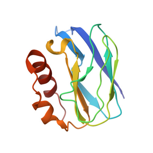
    The crystal structure of native pseudoazurin (HdPAz) from the methylotrophic denitrifying bacterium Hyphomicrobium denitrificans has been determined at a resolution of 1.18 A. After refinement with SHELX employing anisotropic displacement parameters and riding H atoms, R(work) and R(free) were 0.135 and 0.169, respectively. Visualization of the anisotropic displacement parameters as thermal ellipsoids provided insight into the atomic motion within the perturbed type 1 Cu site. The asymmetric unit includes three HdPAz molecules which are tightly packed by head-to-head cupredoxin dimer formation. The shape of the Cu-atom ellipsoid implies significant vibrational motion diagonal to the equatorial xy plane defined by the three ligands (two His and one Cys). The geometric parameters of the type 1 Cu site in the HdPAz structure differ unambiguously from those of other pseudoazurins. It is demonstrated that their structural aspects are consistent with the unique visible absorption spectrum.


  • Organizational Affiliation

    Bioinorganic Chemistry Laboratory, Department of Chemistry, Graduate School of Science, Osaka University, 1-1 Machikaneyama, Toyonaka, Osaka 560-0043, Japan.


Macromolecules
Find similar proteins by:  (by identity cutoff)  |  3D Structure
Entity ID: 1
MoleculeChains Sequence LengthOrganismDetailsImage
Blue copper protein
A, B, C
124Hyphomicrobium denitrificansMutation(s): 0 
UniProt
Find proteins for A7VL37 (Hyphomicrobium denitrificans)
Explore A7VL37 
Go to UniProtKB:  A7VL37
Entity Groups  
Sequence Clusters30% Identity50% Identity70% Identity90% Identity95% Identity100% Identity
UniProt GroupA7VL37
Sequence Annotations
Expand
  • Reference Sequence
Small Molecules
Ligands 2 Unique
IDChains Name / Formula / InChI Key2D Diagram3D Interactions
PO4
Query on PO4

Download Ideal Coordinates CCD File 
E [auth A]
F [auth A]
G [auth A]
H [auth A]
J [auth B]
E [auth A],
F [auth A],
G [auth A],
H [auth A],
J [auth B],
K [auth B],
L [auth B],
M [auth B],
N [auth B],
O [auth B],
Q [auth C],
R [auth C],
S [auth C],
T [auth C],
U [auth C]
PHOSPHATE ION
O4 P
NBIIXXVUZAFLBC-UHFFFAOYSA-K
CU
Query on CU

Download Ideal Coordinates CCD File 
D [auth A],
I [auth B],
P [auth C]
COPPER (II) ION
Cu
JPVYNHNXODAKFH-UHFFFAOYSA-N
Experimental Data & Validation

Experimental Data

  • Method: X-RAY DIFFRACTION
  • Resolution: 1.18 Å
  • R-Value Free: 0.169 
  • R-Value Work: 0.135 
  • R-Value Observed: 0.135 
  • Space Group: C 1 2 1
Unit Cell:
Length ( Å )Angle ( ˚ )
a = 97.69α = 90
b = 50.623β = 90.98
c = 82.378γ = 90
Software Package:
Software NamePurpose
HKL-2000data collection
MOLREPphasing
SHELXL-97refinement
HKL-2000data reduction
SCALEPACKdata scaling

Structure Validation

View Full Validation Report


Entry History 

Deposition Data

Revision History  (Full details and data files)

  • Version 1.0: 2008-12-30
    Type: Initial release
  • Version 1.1: 2011-07-13
    Changes: Version format compliance
  • Version 1.2: 2023-11-01
    Changes: Data collection, Database references, Derived calculations, Refinement description