3EE4

R2-like ligand binding Mn/Fe oxidase from M. tuberculosis


Experimental Data Snapshot

  • Method: X-RAY DIFFRACTION
  • Resolution: 1.90 Å
  • R-Value Free: 0.177 
  • R-Value Work: 0.149 
  • R-Value Observed: 0.150 

wwPDB Validation   3D Report Full Report


Ligand Structure Quality Assessment 


This is version 1.1 of the entry. See complete history


Literature

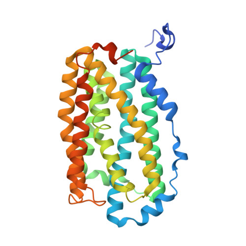
A Mycobacterium tuberculosis ligand-binding Mn/Fe protein reveals a new cofactor in a remodeled R2-protein scaffold

Andersson, C.S.

(2009) Proc Natl Acad Sci U S A 106: 5633-5638

  • DOI: https://doi.org/10.1073/pnas.0812971106
  • Primary Citation of Related Structures:  
    3EE4

  • PubMed Abstract: 

    Chlamydia trachomatis R2c is the prototype for a recently discovered group of ribonucleotide reductase R2 proteins that use a heterodinuclear Mn/Fe redox cofactor for radical generation and storage. Here, we show that the Mycobacterium tuberculosis protein Rv0233, an R2 homologue and a potential virulence factor, contains the heterodinuclear manganese/iron-carboxylate cofactor but displays a drastic remodeling of the R2 protein scaffold into a ligand-binding oxidase. The first structural characterization of the heterodinuclear cofactor shows that the site is highly specific for manganese and iron in their respective positions despite a symmetric arrangement of coordinating residues. In this protein scaffold, the Mn/Fe cofactor supports potent 2-electron oxidations as revealed by an unprecedented tyrosine-valine crosslink in the active site. This wolf in sheep's clothing defines a distinct functional group among R2 homologues and may represent a structural and functional counterpart of the evolutionary ancestor of R2s and bacterial multicomponent monooxygenases.


  • Organizational Affiliation

    Stockholm Center for Biomembrane Research, Department of Biochemistry and Biophysics, Stockholm University, Arrhenius Laboratories for Natural Sciences C4, SE-106 91 Stockholm, Sweden.


Macromolecules
Find similar proteins by:  (by identity cutoff)  |  3D Structure
Entity ID: 1
MoleculeChains Sequence LengthOrganismDetailsImage
Probable ribonucleoside-diphosphate reductase323Mycobacterium tuberculosisMutation(s): 0 
Gene Names: nrdBRv0233
EC: 1.17.4.1
UniProt
Find proteins for P9WH69 (Mycobacterium tuberculosis (strain ATCC 25618 / H37Rv))
Explore P9WH69 
Go to UniProtKB:  P9WH69
Entity Groups  
Sequence Clusters30% Identity50% Identity70% Identity90% Identity95% Identity100% Identity
UniProt GroupP9WH69
Sequence Annotations
Expand
  • Reference Sequence
Experimental Data & Validation

Experimental Data

  • Method: X-RAY DIFFRACTION
  • Resolution: 1.90 Å
  • R-Value Free: 0.177 
  • R-Value Work: 0.149 
  • R-Value Observed: 0.150 
  • Space Group: P 32 2 1
Unit Cell:
Length ( Å )Angle ( ˚ )
a = 54.565α = 90
b = 54.565β = 90
c = 176.651γ = 120
Software Package:
Software NamePurpose
ADSCdata collection
SHARPphasing
REFMACrefinement
MOSFLMdata reduction
SCALAdata scaling

Structure Validation

View Full Validation Report


Ligand Structure Quality Assessment 


Entry History 

Deposition Data

Revision History  (Full details and data files)

  • Version 1.0: 2009-04-07
    Type: Initial release
  • Version 1.1: 2011-07-13
    Changes: Advisory, Version format compliance