3EE2

Structure of human prostaglandin D-synthase (hGSTS1-1) in complex with nocodazole


Experimental Data Snapshot

  • Method: X-RAY DIFFRACTION
  • Resolution: 1.91 Å
  • R-Value Free: 0.232 
  • R-Value Work: 0.183 
  • R-Value Observed: 0.186 

wwPDB Validation   3D Report Full Report


Ligand Structure Quality Assessment 


This is version 1.4 of the entry. See complete history


Literature

Identification and characterisation of new inhibitors for the human hematopoietic prostaglandin D(2) synthase

Weber, J.E.Oakley, A.J.Christ, A.N.Clark, A.G.Hayes, J.D.Hall, R.Hume, D.A.Board, P.G.Smythe, M.L.Flanagan, J.U.

(2010) Eur J Med Chem 45: 447-454

  • DOI: https://doi.org/10.1016/j.ejmech.2009.10.025
  • Primary Citation of Related Structures:  
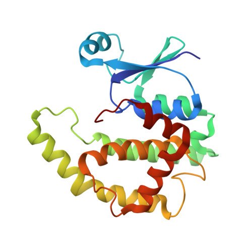
    3EE2

  • PubMed Abstract: 

    Prostaglandin D(2) synthesised by the hematopoietic prostaglandin D(2) synthase has a pro-inflammatory effect in allergic asthma, regulating many hallmark characteristics of the disease. Here we describe identification of hematopoietic prostaglandin D(2) synthase inhibitors including cibacron blue, bromosulfophthalein and ethacrynic acid. Expansion around the drug-like ethacrynic acid identified a novel inhibitor, nocodazole, and a fragment representing its aromatic core. Nocodazole binding was further characterised by docking calculations in combination with conformational strain analysis. The benzyl thiophene core was predicted to be buried in the active site, binding in the putative prostaglandin binding site, and a likely hydrogen bond donor site identified. X-ray crystallographic studies supported the predicted binding mode.


  • Organizational Affiliation

    The University of Queensland, Institute for Molecular Bioscience, Building 80, St Lucia, Queensland 4072, Australia.


Macromolecules
Find similar proteins by:  (by identity cutoff)  |  3D Structure
Entity ID: 1
MoleculeChains Sequence LengthOrganismDetailsImage
Glutathione-requiring prostaglandin D synthase
A, B
199Homo sapiensMutation(s): 0 
Gene Names: PGDS
EC: 5.3.99.2
UniProt & NIH Common Fund Data Resources
Find proteins for O60760 (Homo sapiens)
Explore O60760 
Go to UniProtKB:  O60760
PHAROS:  O60760
GTEx:  ENSG00000163106 
Entity Groups  
Sequence Clusters30% Identity50% Identity70% Identity90% Identity95% Identity100% Identity
UniProt GroupO60760
Sequence Annotations
Expand
  • Reference Sequence
Small Molecules
Binding Affinity Annotations 
IDSourceBinding Affinity
NZO PDBBind:  3EE2 IC50: 6.50e+4 (nM) from 1 assay(s)
Binding MOAD:  3EE2 IC50: 6.50e+4 (nM) from 1 assay(s)
Experimental Data & Validation

Experimental Data

  • Method: X-RAY DIFFRACTION
  • Resolution: 1.91 Å
  • R-Value Free: 0.232 
  • R-Value Work: 0.183 
  • R-Value Observed: 0.186 
  • Space Group: P 21 21 21
Unit Cell:
Length ( Å )Angle ( ˚ )
a = 47.22α = 90
b = 79.359β = 90
c = 107.547γ = 90
Software Package:
Software NamePurpose
ADSCdata collection
MOLREPphasing
REFMACrefinement
HKL-2000data reduction
HKL-2000data scaling

Structure Validation

View Full Validation Report


Ligand Structure Quality Assessment 


Entry History 

Deposition Data

  • Released Date: 2008-09-16 
  • Deposition Author(s): Oakley, A.J.

Revision History  (Full details and data files)

  • Version 1.0: 2008-09-16
    Type: Initial release
  • Version 1.1: 2011-07-13
    Changes: Advisory, Version format compliance
  • Version 1.2: 2011-12-14
    Changes: Non-polymer description
  • Version 1.3: 2017-10-25
    Changes: Refinement description
  • Version 1.4: 2023-11-01
    Changes: Data collection, Database references, Derived calculations, Refinement description