3ECY

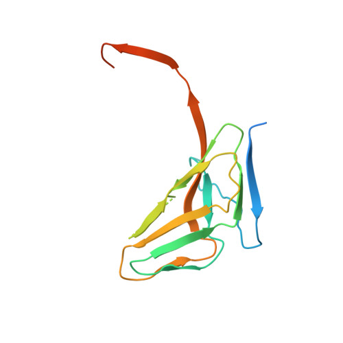
Crystal structural analysis of Drosophila melanogaster dUTPase


Experimental Data Snapshot

  • Method: X-RAY DIFFRACTION
  • Resolution: 1.88 Å
  • R-Value Free: 0.222 
  • R-Value Work: 0.200 
  • R-Value Observed: 0.202 

wwPDB Validation   3D Report Full Report


This is version 1.2 of the entry. See complete history


Literature

Molecular shape and prominent role of beta-strand swapping in organization of dUTPase oligomers.

Takacs, E.Barabas, O.Petoukhov, M.V.Svergun, D.I.Vertessy, B.G.

(2009) FEBS Lett 583: 865-871

  • DOI: https://doi.org/10.1016/j.febslet.2009.02.011
  • Primary Citation of Related Structures:  
    3ECY

  • PubMed Abstract: 

    Most dUTP pyrophosphatases (dUTPases) are homotrimers with interfaces formed between subunit surfaces, in the central channel, and by C-terminal beta-strand swapping. Analysis of intersubunit interactions reveals an important cohesive role for the C-terminus. This is reflected in the crystal structure of fruitfly dUTPase displaying a dimeric organization in crystals grown in alcohol solution, where only beta-strand swapping interactions between subunits are retained from the usual trimer structure. Mutations of a suggested hinge proline destabilize human and Escherichia coli dUTPases without preventing trimeric organization. Trimer formation was, however, prevented in the human enzyme by truncating the C-terminus before the swapping arm. The molecular shape of full-length enzymes in solution reveals the localization and variation in flexibility of N- and C-terminal segments.


  • Organizational Affiliation

    Institute of Enzimology, BRC, Hungarian Academy of Sciences, Budapest, Hungary.


Macromolecules
Find similar proteins by:  (by identity cutoff)  |  3D Structure
Entity ID: 1
MoleculeChains Sequence LengthOrganismDetailsImage
CG4584-PA, isoform A (BcDNA.LD08534)
A, B
160Drosophila melanogasterMutation(s): 0 
Gene Names: dUTPaseBcDNA.LD08534CG4584Dmel_CG4584
EC: 3.6.1.23
UniProt
Find proteins for Q9V3I1 (Drosophila melanogaster)
Explore Q9V3I1 
Go to UniProtKB:  Q9V3I1
Entity Groups  
Sequence Clusters30% Identity50% Identity70% Identity90% Identity95% Identity100% Identity
UniProt GroupQ9V3I1
Sequence Annotations
Expand
  • Reference Sequence
Experimental Data & Validation

Experimental Data

  • Method: X-RAY DIFFRACTION
  • Resolution: 1.88 Å
  • R-Value Free: 0.222 
  • R-Value Work: 0.200 
  • R-Value Observed: 0.202 
  • Space Group: P 41
Unit Cell:
Length ( Å )Angle ( ˚ )
a = 42.799α = 90
b = 42.799β = 90
c = 169.384γ = 90
Software Package:
Software NamePurpose
REFMACrefinement
XDSdata reduction
XSCALEdata scaling
MOLREPphasing

Structure Validation

View Full Validation Report


Entry History 

Deposition Data

Revision History  (Full details and data files)

  • Version 1.0: 2008-10-07
    Type: Initial release
  • Version 1.1: 2011-07-13
    Changes: Advisory, Version format compliance
  • Version 1.2: 2023-08-30
    Changes: Data collection, Database references, Refinement description