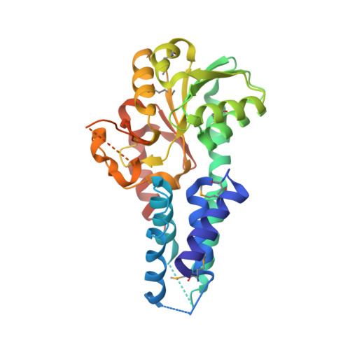
Crystal structure of the alpha subunit of human translation initiation factor 2B
Hiyama, T.B., Ito, T., Imataka, H., Yokoyama, S.(2009) J Mol Biol 392: 937-951
- PubMed: 19631657
- DOI: https://doi.org/10.1016/j.jmb.2009.07.054
- Primary Citation of Related Structures:
3ECS - PubMed Abstract:
Eukaryotic translation initiation factor 2B (eIF2B) is the heteropentameric guanine-nucleotide exchange factor specific for eukaryotic initiation factor 2 (eIF2). Under stressed conditions, guanine-nucleotide exchange is strongly inhibited by the tight binding of phosphorylated eIF2 to eIF2B. Here, we report the crystal structure of the alpha subunit of human eIF2B at 2.65 A resolution. The eIF2Balpha structure consists of the N-terminal alpha-helical domain and the C-terminal Rossmann-fold-like domain. A positively charged pocket, whose entrance is about 15-17 A in diameter, resides at the boundary between the two domains. A sulfate ion is located at the bottom of the pocket (about 16 A in depth). The residues comprising the sulfate-ion-binding site are strictly conserved in eIF2Balpha. Since this deep, wide pocket with the sulfate-ion-binding site is not conserved in distant homologues, including 5-methylthioribose 1-phosphate isomerases, these characteristics may be distinctive of eIF2Balpha. Interestingly, the yeast eIF2Balpha missense mutations that reduce the eIF2B sensitivity to phosphorylated eIF2 are mapped on the other side of the pocket. One of the three human eIF2Balpha missense mutations that induce the lethal brain disorder vanishing white matter or childhood ataxia with central nervous system hypomyelination is mapped inside the pocket. The beta and delta subunits of eIF2B are homologous to eIF2Balpha and may have tertiary structures similar to the present eIF2Balpha structure. The sulfate-ion-binding residues of eIF2Balpha are well conserved in eIF2Bbeta/delta. The abovementioned yeast and human missense mutations of eIF2Bbeta/delta were also mapped on the eIF2Balpha structure, which revealed that the human mutations are clustered on the same side as the pocket, while the yeast mutations reside on the opposite side. As most of the mutated residues are exposed on the surface of the eIF2B subunit structure, these exposed residues are likely to be involved in either the subunit interactions or the interaction with eIF2.
Organizational Affiliation:
Department of Biophysics and Biochemistry, Graduate School of Science, the University of Tokyo, 7-3-1 Hongo, Bunkyo-ku, Tokyo 113-0033, Japan.