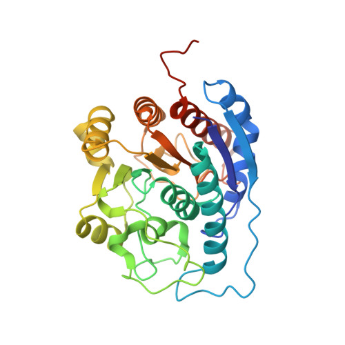
Probing the specificity determinants of amino acid recognition by arginase.
Shishova, E.Y., Di Costanzo, L., Emig, F.A., Ash, D.E., Christianson, D.W.(2009) Biochemistry 48: 121-131
- PubMed: 19093830
- DOI: https://doi.org/10.1021/bi801911v
- Primary Citation of Related Structures:
3E6K, 3E6V, 3E8Q, 3E8Z, 3E9B - PubMed Abstract:
Arginase is a binuclear manganese metalloenzyme that serves as a therapeutic target for the treatment of asthma, erectile dysfunction, and atherosclerosis. In order to better understand the molecular basis of inhibitor affinity, we have employed site-directed mutagenesis, enzyme kinetics, and X-ray crystallography to probe the molecular recognition of the amino acid moiety (i.e., the alpha-amino and alpha-carboxylate groups) of substrate l-arginine and inhibitors in the active site of arginase I. Specifically, we focus on (1) a water-mediated hydrogen bond between the substrate alpha-carboxylate and T135, (2) a direct hydrogen bond between the substrate alpha-carboxylate and N130, and (3) a direct charged hydrogen bond between the substrate alpha-amino group and D183. Amino acid substitutions for T135, N130, and D183 generally compromise substrate affinity as reflected by increased K(M) values but have less pronounced effects on catalytic function as reflected by minimal variations of k(cat). As with substrate K(M) values, inhibitor K(d) values increase for binding to enzyme mutants and suggest that the relative contribution of intermolecular interactions to amino acid affinity in the arginase active site is water-mediated hydrogen bond < direct hydrogen bond < direct charged hydrogen bond. Structural comparisons of arginase with the related binuclear manganese metalloenzymes agmatinase and proclavaminic acid amidinohydrolase suggest that the evolution of substrate recognition in the arginase fold occurs by mutation of residues contained in specificity loops flanking the mouth of the active site (especially loops 4 and 5), thereby allowing diverse guanidinium substrates to be accommodated for catalysis.
Organizational Affiliation:
Roy and Diana Vagelos Laboratories, Department of Chemistry, University of Pennsylvania, Philadelphia, Pennsylvania 19104-6323, USA.