3E6J

Crystal Structure of Variable Lymphocyte Receptor (VLR) RBC36 in Complex with H-trisaccharide


Experimental Data Snapshot

  • Method: X-RAY DIFFRACTION
  • Resolution: 1.67 Å
  • R-Value Free: 0.182 
  • R-Value Work: 0.146 
  • R-Value Observed: 0.148 

wwPDB Validation   3D Report Full Report


This is version 2.1 of the entry. See complete history


Literature

Antigen recognition by variable lymphocyte receptors.

Han, B.W.Herrin, B.R.Cooper, M.D.Wilson, I.A.

(2008) Science 321: 1834-1837

  • DOI: https://doi.org/10.1126/science.1162484
  • Primary Citation of Related Structures:  
    3E6J

  • PubMed Abstract: 

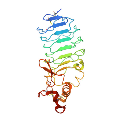
    Variable lymphocyte receptors (VLRs) rather than antibodies play the primary role in recognition of antigens in the adaptive immune system of jawless vertebrates. Combinatorial assembly of leucine-rich repeat (LRR) gene segments achieves the required repertoire for antigen recognition. We have determined a crystal structure for a VLR-antigen complex, VLR RBC36 in complex with the H-antigen trisaccharide from human blood type O erythrocytes, at 1.67 angstrom resolution. RBC36 binds the H-trisaccharide on the concave surface of the LRR modules of the solenoid structure where three key hydrophilic residues, multiple van der Waals interactions, and the highly variable insert of the carboxyl-terminal LRR module determine antigen recognition and specificity. The concave surface assembled from the most highly variable regions of the LRRs, along with diversity in the sequence and length of the highly variable insert, can account for the recognition of diverse antigens by VLRs.


  • Organizational Affiliation

    Department of Molecular Biology, Scripps Research Institute, La Jolla, CA 92037, USA.


Macromolecules
Find similar proteins by:  (by identity cutoff)  |  3D Structure
Entity ID: 1
MoleculeChains Sequence LengthOrganismDetailsImage
Variable lymphocyte receptor diversity region229Petromyzon marinusMutation(s): 0 
Gene Names: VLR
UniProt
Find proteins for Q2VGE8 (Petromyzon marinus)
Explore Q2VGE8 
Go to UniProtKB:  Q2VGE8
Entity Groups  
Sequence Clusters30% Identity50% Identity70% Identity90% Identity95% Identity100% Identity
UniProt GroupQ2VGE8
Sequence Annotations
Expand
  • Reference Sequence
Oligosaccharides

Help

Entity ID: 2
MoleculeChains Length2D Diagram Glycosylation3D Interactions
alpha-L-fucopyranose-(1-2)-beta-D-galactopyranose-(1-4)-2-acetamido-2-deoxy-beta-D-glucopyranose
B
3N/A
Glycosylation Resources
GlyTouCan:  G61459JW
GlyCosmos:  G61459JW
GlyGen:  G61459JW
Biologically Interesting Molecules (External Reference) 1 Unique
Experimental Data & Validation

Experimental Data

  • Method: X-RAY DIFFRACTION
  • Resolution: 1.67 Å
  • R-Value Free: 0.182 
  • R-Value Work: 0.146 
  • R-Value Observed: 0.148 
  • Space Group: P 1 21 1
Unit Cell:
Length ( Å )Angle ( ˚ )
a = 54.221α = 90
b = 48.564β = 116.51
c = 55.477γ = 90
Software Package:
Software NamePurpose
REFMACrefinement
HKL-2000data collection
HKL-2000data reduction
HKL-2000data scaling
MOLREPphasing

Structure Validation

View Full Validation Report


Entry History 

Deposition Data

Revision History  (Full details and data files)

  • Version 1.0: 2008-10-07
    Type: Initial release
  • Version 1.1: 2011-07-13
    Changes: Non-polymer description, Version format compliance
  • Version 2.0: 2020-07-29
    Type: Remediation
    Reason: Carbohydrate remediation
    Changes: Advisory, Atomic model, Data collection, Database references, Derived calculations, Non-polymer description, Structure summary
  • Version 2.1: 2023-08-30
    Changes: Data collection, Database references, Refinement description, Structure summary