3E17

Crystal structure of the second PDZ domain from human Zona Occludens-2


Experimental Data Snapshot

  • Method: X-RAY DIFFRACTION
  • Resolution: 1.75 Å
  • R-Value Free: 0.237 
  • R-Value Work: 0.206 
  • R-Value Observed: 0.208 

wwPDB Validation   3D Report Full Report


This is version 1.3 of the entry. See complete history


Literature

Structure of the second PDZ domain from human zonula occludens 2

Chen, H.Tong, S.Li, X.Wu, J.Zhu, Z.Niu, L.Teng, M.

(2009) Acta Crystallogr Sect F Struct Biol Cryst Commun 65: 327-330

  • DOI: https://doi.org/10.1107/S1744309109002334
  • Primary Citation of Related Structures:  
    3E17

  • PubMed Abstract: 

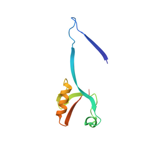
    Human zonula occludens 2 (ZO-2) protein is a multi-domain protein that consists of an SH3 domain, a GK domain and three copies of a PDZ domain with slight divergence. The three PDZ domains act as protein-recognition modules that may mediate protein assembly and subunit localization. The crystal structure of the second PDZ domain of ZO-2 (ZO-2 PDZ2) was determined by molecular replacement at 1.75 A resolution, revealing a dimer in the asymmetric unit. The dimer is stabilized by extensive symmetrical domain-swapping of the beta1 and beta2 strands. Structural comparison shows that the ZO-2 PDZ2 homodimer may have a similar ligand-binding pattern to the ZO-1 PDZ2-connexin 43 complex.


  • Organizational Affiliation

    Hefei National Laboratory for Physical Sciences at Microscale, School of Life Sciences, University of Science and Technology of China, Hefei, Anhui, People's Republic of China.


Macromolecules
Find similar proteins by:  (by identity cutoff)  |  3D Structure
Entity ID: 1
MoleculeChains Sequence LengthOrganismDetailsImage
Tight junction protein ZO-2
A, B
88Homo sapiensMutation(s): 0 
Gene Names: TJP2X104ZO2
UniProt & NIH Common Fund Data Resources
Find proteins for Q9UDY2 (Homo sapiens)
Explore Q9UDY2 
Go to UniProtKB:  Q9UDY2
PHAROS:  Q9UDY2
GTEx:  ENSG00000119139 
Entity Groups  
Sequence Clusters30% Identity50% Identity70% Identity90% Identity95% Identity100% Identity
UniProt GroupQ9UDY2
Sequence Annotations
Expand
  • Reference Sequence
Experimental Data & Validation

Experimental Data

  • Method: X-RAY DIFFRACTION
  • Resolution: 1.75 Å
  • R-Value Free: 0.237 
  • R-Value Work: 0.206 
  • R-Value Observed: 0.208 
  • Space Group: P 1
Unit Cell:
Length ( Å )Angle ( ˚ )
a = 30.177α = 80.05
b = 41.325β = 68.63
c = 41.292γ = 68.52
Software Package:
Software NamePurpose
DENZOdata reduction
SCALEPACKdata scaling
MOLREPphasing
REFMACrefinement
PDB_EXTRACTdata extraction
MAR345dtbdata collection
AUTOMARdata reduction

Structure Validation

View Full Validation Report


Entry History 

Deposition Data

Revision History  (Full details and data files)

  • Version 1.0: 2009-07-21
    Type: Initial release
  • Version 1.1: 2011-07-13
    Changes: Version format compliance
  • Version 1.2: 2017-10-25
    Changes: Refinement description
  • Version 1.3: 2023-11-01
    Changes: Data collection, Database references, Refinement description