


The crystal structure of a conserved domain from a protein of Geobacter sulfurreducens PCA
Tan, K., Bigelow, L., Clancy, S., Joachimiak, A.To be published.
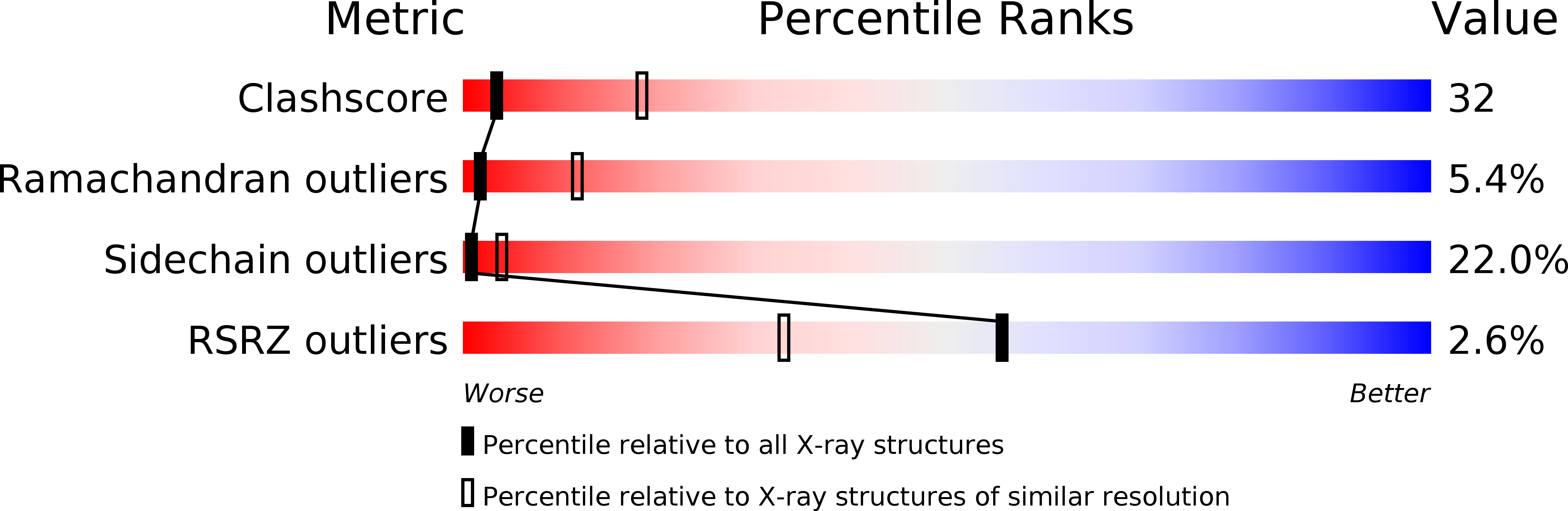
Experimental Data Snapshot
wwPDB Validation 3D Report Full Report
Entity ID: 1 | |||||
|---|---|---|---|---|---|
| Molecule | Chains | Sequence Length | Organism | Details | Image |
| Conserved domain protein | 181 | Geobacter sulfurreducens | Mutation(s): 0 Gene Names: GSU1116 | 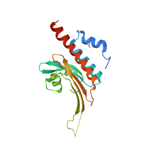 | |
UniProt | |||||
Find proteins for Q74E48 (Geobacter sulfurreducens (strain ATCC 51573 / DSM 12127 / PCA)) Explore Q74E48 Go to UniProtKB: Q74E48 | |||||
Entity Groups | |||||
| Sequence Clusters | 30% Identity50% Identity70% Identity90% Identity95% Identity100% Identity | ||||
| UniProt Group | Q74E48 | ||||
Sequence AnnotationsExpand | |||||
| |||||
| Ligands 1 Unique | |||||
|---|---|---|---|---|---|
| ID | Chains | Name / Formula / InChI Key | 2D Diagram | 3D Interactions | |
| NA Query on NA | C [auth A], D [auth B] | SODIUM ION Na FKNQFGJONOIPTF-UHFFFAOYSA-N |  | ||
| Modified Residues 1 Unique | |||||
|---|---|---|---|---|---|
| ID | Chains | Type | Formula | 2D Diagram | Parent |
| MSE Query on MSE | A, B | L-PEPTIDE LINKING | C5 H11 N O2 Se |  | MET |
| Length ( Å ) | Angle ( ˚ ) |
|---|---|
| a = 59.421 | α = 90 |
| b = 59.421 | β = 90 |
| c = 223.175 | γ = 120 |
| Software Name | Purpose |
|---|---|
| REFMAC | refinement |
| SBC-Collect | data collection |
| HKL-3000 | data reduction |
| HKL-3000 | data scaling |
| SHELXD | phasing |
| MLPHARE | phasing |
| DM | phasing |
| RESOLVE | phasing |
| HKL-3000 | phasing |
RCSB PDB (citation) is hosted by
RCSB PDB is a member of the

