Genetic and structural insights into the dissemination potential of the extremely broad-spectrum class A beta-lactamase KPC-2 identified in an Escherichia coli strain and an Enterobacter cloacae strain isolated from the same patient in France.
Petrella, S., Ziental-Gelus, N., Mayer, C., Renard, M., Jarlier, V., Sougakoff, W.(2008) Antimicrob Agents Chemother 52: 3725-3736
- PubMed: 18625772
- DOI: https://doi.org/10.1128/AAC.00163-08
- Primary Citation of Related Structures:
3DW0 - PubMed Abstract:
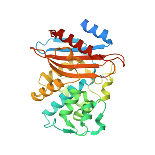
Two clinical strains of Escherichia coli (2138) and Enterobacter cloacae (7506) isolated from the same patient in France and showing resistance to extended-spectrum cephalosporins and low susceptibility to imipenem were investigated. Both strains harbored the plasmid-contained bla(TEM-1) and bla(KPC-2) genes. bla(KLUC-2), encoding a mutant of the chromosomal beta-lactamase of Kluyvera cryocrescens, was also identified at a plasmid location in E. cloacae 7506, suggesting the ISEcp1-assisted escape of bla(KLUC) from the chromosome. Determination of the KPC-2 structure at 1.6 A revealed that the binding site was occupied by the C-terminal (C-ter) residues coming from a symmetric KPC-2 monomer, with the ultimate C-ter Glu interacting with Ser130, Lys234, Thr235, and Thr237 in the active site. This mode of binding can be paralleled to the inhibition of the TEM-1 beta-lactamase by the inhibitory protein BLIP. Determination of the 1.23-A structure of a KPC-2 mutant in which the five C-ter residues were deleted revealed that the catalytic site was filled by a citrate molecule. Structure analysis and docking simulations with cefotaxime and imipenem provided further insights into the molecular basis of the extremely broad spectrum of KPC-2, which behaves as a cefotaximase with significant activity against carbapenems. In particular, residues 104, 105, 132, and 167 draw a binding cavity capable of accommodating both the aminothiazole moiety of cefotaxime and the 6 alpha-hydroxyethyl group of imipenem, with the binding of the former drug being also favored by a significant degree of freedom at the level of the loop at positions 96 to 105 and by an enlargement of the binding site at the end of strand beta 3.
Organizational Affiliation:
UPMC Univ Paris 06, EA1541, Bacteriologie-Hygiène, Paris, France.