3DUY

Crystal structure of human beta-secretase in complex with NVP-AFJ144


Experimental Data Snapshot

  • Method: X-RAY DIFFRACTION
  • Resolution: 1.97 Å
  • R-Value Free: 0.223 
  • R-Value Work: 0.202 
  • R-Value Observed: 0.204 

wwPDB Validation   3D Report Full Report


Ligand Structure Quality Assessment 


This is version 1.3 of the entry. See complete history


Literature

Structure-based design and synthesis of macrocyclic peptidomimetic beta-secretase (BACE-1) inhibitors.

Machauer, R.Veenstra, S.Rondeau, J.M.Tintelnot-Blomley, M.Betschart, C.Neumann, U.Paganetti, P.

(2009) Bioorg Med Chem Lett 19: 1361-1365

  • DOI: https://doi.org/10.1016/j.bmcl.2009.01.036
  • Primary Citation of Related Structures:  
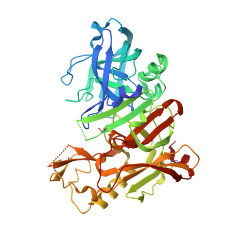
    3DUY

  • PubMed Abstract: 

    The hydroxyethylene octapeptide inhibitor OM99-2 served as starting point to create the tripeptide inhibitor 1 and its analogues 2a and b. An X-ray co-crystal structure of 1 with BACE-1 allowed the design and syntheses of a series of macrocyclic analogues 3a-h covalently linking the P1 and P3 side-chains. These inhibitors show improved enzymatic potency over their open-chain analogue. Inhibitor 3h also shows activity in a cellular system.


  • Organizational Affiliation

    Novartis Institutes for BioMedical Research, Novartis Pharma AG, PO Box, CH-4002 Basel, Switzerland.


Macromolecules
Find similar proteins by:  (by identity cutoff)  |  3D Structure
Entity ID: 1
MoleculeChains Sequence LengthOrganismDetailsImage
Beta-secretase 1
A, B, C
402Homo sapiensMutation(s): 0 
Gene Names: BACE1BACE
EC: 3.4.23.46
UniProt & NIH Common Fund Data Resources
Find proteins for P56817 (Homo sapiens)
Explore P56817 
Go to UniProtKB:  P56817
PHAROS:  P56817
GTEx:  ENSG00000186318 
Entity Groups  
Sequence Clusters30% Identity50% Identity70% Identity90% Identity95% Identity100% Identity
UniProt GroupP56817
Sequence Annotations
Expand
  • Reference Sequence
Small Molecules
Ligands 1 Unique
IDChains Name / Formula / InChI Key2D Diagram3D Interactions
AFJ
Query on AFJ

Download Ideal Coordinates CCD File 
D [auth A],
E [auth B],
F [auth C]
(2R,4S,5S)-N-butyl-4-hydroxy-2,7-dimethyl-5-{[N-(4-methylpentanoyl)-L-methionyl]amino}octanamide
C25 H49 N3 O4 S
XTRMRNMVFXWTCC-MLNNCEHLSA-N
Binding Affinity Annotations 
IDSourceBinding Affinity
AFJ Binding MOAD:  3DUY IC50: 1400 (nM) from 1 assay(s)
PDBBind:  3DUY IC50: 1400 (nM) from 1 assay(s)
BindingDB:  3DUY IC50: min: 1400, max: 1.00e+4 (nM) from 2 assay(s)
Experimental Data & Validation

Experimental Data

  • Method: X-RAY DIFFRACTION
  • Resolution: 1.97 Å
  • R-Value Free: 0.223 
  • R-Value Work: 0.202 
  • R-Value Observed: 0.204 
  • Space Group: P 1 21 1
Unit Cell:
Length ( Å )Angle ( ˚ )
a = 82.537α = 90
b = 103.062β = 103.64
c = 100.602γ = 90
Software Package:
Software NamePurpose
XSCALEdata scaling
CNSrefinement
PDB_EXTRACTdata extraction
XDSdata reduction
CNXphasing
CNXrefinement

Structure Validation

View Full Validation Report


Ligand Structure Quality Assessment 


Entry History 

Deposition Data

Revision History  (Full details and data files)

  • Version 1.0: 2009-02-24
    Type: Initial release
  • Version 1.1: 2011-07-13
    Changes: Version format compliance
  • Version 1.2: 2017-10-25
    Changes: Refinement description
  • Version 1.3: 2023-08-30
    Changes: Data collection, Database references, Derived calculations, Refinement description