3DTU

Catalytic core subunits (I and II) of cytochrome c oxidase from Rhodobacter sphaeroides complexed with deoxycholic acid


Experimental Data Snapshot

  • Method: X-RAY DIFFRACTION
  • Resolution: 2.15 Å
  • R-Value Free: 0.212 
  • R-Value Work: 0.184 
  • R-Value Observed: 0.184 

wwPDB Validation   3D Report Full Report


Ligand Structure Quality Assessment 


This is version 1.3 of the entry. See complete history


Literature

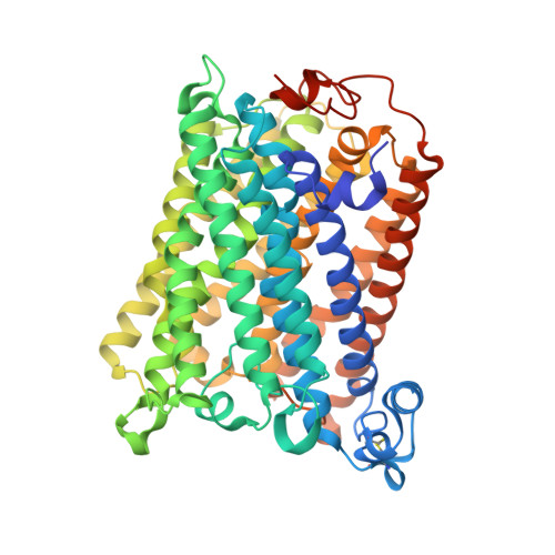
A conserved steroid binding site in cytochrome C oxidase.

Qin, L.Mills, D.A.Buhrow, L.Hiser, C.Ferguson-Miller, S.

(2008) Biochemistry 47: 9931-9933

  • DOI: https://doi.org/10.1021/bi8013483
  • Primary Citation of Related Structures:  
    3DTU

  • PubMed Abstract: 

    Micromolar concentrations of the bile salt deoxycholate are shown to rescue the activity of an inactive mutant, E101A, in the K proton pathway of Rhodobacter sphaeroides cytochrome c oxidase. A crystal structure of the wild-type enzyme reveals, as predicted, deoxycholate bound with its carboxyl group at the entrance of the K path. Since cholate is a known potent inhibitor of bovine oxidase and is seen in a similar position in the bovine structure, the crystallographically defined, conserved steroid binding site could reveal a regulatory site for steroids or structurally related molecules that act on the essential K proton path.


  • Organizational Affiliation

    Biochemistry and Molecular Biology Department, Michigan State University, East Lansing, Michigan 48824, USA.


Macromolecules
Find similar proteins by:  (by identity cutoff)  |  3D Structure
Entity ID: 1
MoleculeChains Sequence LengthOrganismDetailsImage
Cytochrome c oxidase subunit 1
A, C
566Cereibacter sphaeroidesMutation(s): 0 
Gene Names: ctaD
EC: 1.9.3.1
Membrane Entity: Yes 
UniProt
Find proteins for P33517 (Cereibacter sphaeroides)
Explore P33517 
Go to UniProtKB:  P33517
Entity Groups  
Sequence Clusters30% Identity50% Identity70% Identity90% Identity95% Identity100% Identity
UniProt GroupP33517
Sequence Annotations
Expand
  • Reference Sequence
Find similar proteins by:  (by identity cutoff)  |  3D Structure
Entity ID: 2
MoleculeChains Sequence LengthOrganismDetailsImage
Cytochrome c oxidase subunit 2
B, D
262Cereibacter sphaeroidesMutation(s): 0 
Gene Names: ctaCcoxIIctaB
EC: 1.9.3.1
Membrane Entity: Yes 
UniProt
Find proteins for Q03736 (Cereibacter sphaeroides)
Explore Q03736 
Go to UniProtKB:  Q03736
Entity Groups  
Sequence Clusters30% Identity50% Identity70% Identity90% Identity95% Identity100% Identity
UniProt GroupQ03736
Sequence Annotations
Expand
  • Reference Sequence
Small Molecules
Ligands 11 Unique
IDChains Name / Formula / InChI Key2D Diagram3D Interactions
HEA
Query on HEA

Download Ideal Coordinates CCD File 
N [auth A],
O [auth A],
PA [auth C],
QA [auth C]
HEME-A
C49 H56 Fe N4 O6
ZGGYGTCPXNDTRV-PRYGPKJJSA-L
DMU
Query on DMU

Download Ideal Coordinates CCD File 
AB [auth D]
E [auth A]
F [auth A]
G [auth A]
H [auth A]
AB [auth D],
E [auth A],
F [auth A],
G [auth A],
H [auth A],
IA [auth C],
JA [auth C],
X [auth B],
Y [auth B]
DECYL-BETA-D-MALTOPYRANOSIDE
C22 H42 O11
WOQQAWHSKSSAGF-WXFJLFHKSA-N
DXC
Query on DXC

Download Ideal Coordinates CCD File 
RA [auth C](3ALPHA,5BETA,12ALPHA)-3,12-DIHYDROXYCHOLAN-24-OIC ACID
C24 H40 O4
KXGVEGMKQFWNSR-LLQZFEROSA-N
TRD
Query on TRD

Download Ideal Coordinates CCD File 
EA [auth B]
FA [auth B]
FB [auth D]
GA [auth B]
GB [auth D]
EA [auth B],
FA [auth B],
FB [auth D],
GA [auth B],
GB [auth D],
HB [auth D],
IB [auth D],
P [auth A],
Q [auth A],
R [auth A],
S [auth A],
SA [auth C],
T [auth A],
TA [auth C],
U [auth A],
UA [auth C],
V [auth A],
VA [auth C],
W [auth A],
WA [auth C],
XA [auth C],
YA [auth C],
ZA [auth C]
TRIDECANE
C13 H28
IIYFAKIEWZDVMP-UHFFFAOYSA-N
HTO
Query on HTO

Download Ideal Coordinates CCD File 
HA [auth B]HEPTANE-1,2,3-TRIOL
C7 H16 O3
HXYCHJFUBNTKQR-RNFRBKRXSA-N
CD
Query on CD

Download Ideal Coordinates CCD File 
BA [auth B],
CA [auth B],
DA [auth B],
DB [auth D],
EB [auth D]
CADMIUM ION
Cd
WLZRMCYVCSSEQC-UHFFFAOYSA-N
PO4
Query on PO4

Download Ideal Coordinates CCD File 
M [auth A],
OA [auth C]
PHOSPHATE ION
O4 P
NBIIXXVUZAFLBC-UHFFFAOYSA-K
CU
Query on CU

Download Ideal Coordinates CCD File 
AA [auth B]
BB [auth D]
CB [auth D]
I [auth A]
KA [auth C]
AA [auth B],
BB [auth D],
CB [auth D],
I [auth A],
KA [auth C],
Z [auth B]
COPPER (II) ION
Cu
JPVYNHNXODAKFH-UHFFFAOYSA-N
CA
Query on CA

Download Ideal Coordinates CCD File 
K [auth A],
MA [auth C]
CALCIUM ION
Ca
BHPQYMZQTOCNFJ-UHFFFAOYSA-N
MG
Query on MG

Download Ideal Coordinates CCD File 
J [auth A],
LA [auth C]
MAGNESIUM ION
Mg
JLVVSXFLKOJNIY-UHFFFAOYSA-N
OH
Query on OH

Download Ideal Coordinates CCD File 
L [auth A],
NA [auth C]
HYDROXIDE ION
H O
XLYOFNOQVPJJNP-UHFFFAOYSA-M
Experimental Data & Validation

Experimental Data

  • Method: X-RAY DIFFRACTION
  • Resolution: 2.15 Å
  • R-Value Free: 0.212 
  • R-Value Work: 0.184 
  • R-Value Observed: 0.184 
  • Space Group: P 21 21 21
Unit Cell:
Length ( Å )Angle ( ˚ )
a = 123.243α = 90
b = 132.052β = 90
c = 167.966γ = 90
Software Package:
Software NamePurpose
DENZOdata reduction
SCALEPACKdata scaling
REFMACrefinement
PDB_EXTRACTdata extraction
HKL-2000data collection
PHASERphasing

Structure Validation

View Full Validation Report


Ligand Structure Quality Assessment 


Entry History 

Deposition Data

Revision History  (Full details and data files)

  • Version 1.0: 2008-09-16
    Type: Initial release
  • Version 1.1: 2011-07-13
    Changes: Version format compliance
  • Version 1.2: 2017-10-25
    Changes: Advisory, Refinement description
  • Version 1.3: 2023-08-30
    Changes: Advisory, Data collection, Database references, Derived calculations, Refinement description