3DSL

The Three-dimensional Structure of Bothropasin, the Main Hemorrhagic Factor from Bothrops jararaca venom.


Experimental Data Snapshot

  • Method: X-RAY DIFFRACTION
  • Resolution: 2.70 Å
  • R-Value Free: 0.294 
  • R-Value Work: 0.210 
  • R-Value Observed: 0.217 

wwPDB Validation   3D Report Full Report


Ligand Structure Quality Assessment 


This is version 1.2 of the entry. See complete history


Literature

The three-dimensional structure of bothropasin, the main hemorrhagic factor from Bothrops jararaca venom: Insights for a new classification of snake venom metalloprotease subgroups.

Muniz, J.R.Ambrosio, A.L.Selistre-de-Araujo, H.S.Cominetti, M.R.Moura-da-Silva, A.M.Oliva, G.Garratt, R.C.Souza, D.H.

(2008) Toxicon 52: 807-816

  • DOI: https://doi.org/10.1016/j.toxicon.2008.08.021
  • Primary Citation of Related Structures:  
    3DSL

  • PubMed Abstract: 

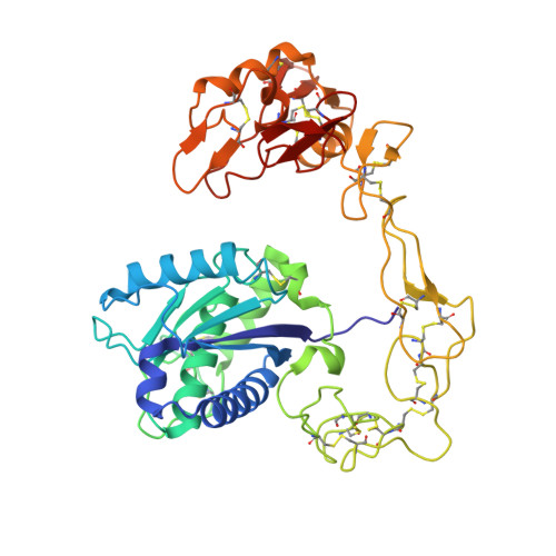
    Bothropasin is a 48kDa hemorrhagic PIII snake venom metalloprotease (SVMP) isolated from Bothrops jararaca, containing disintegrin/cysteine-rich adhesive domains. Here we present the crystal structure of bothropasin complexed with the inhibitor POL647. The catalytic domain consists of a scaffold of two subdomains organized similarly to those described for other SVMPs, including the zinc and calcium-binding sites. The free cysteine residue Cys189 is located within a hydrophobic core and it is not available for disulfide bonding or other interactions. There is no identifiable secondary structure for the disintegrin domain, but instead it is composed mostly of loops stabilized by seven disulfide bonds and by two calcium ions. The ECD region is in a loop and is structurally related to the RGD region of RGD disintegrins, which are derived from PII SVMPs. The ECD motif is stabilized by the Cys277-Cys310 disulfide bond (between the disintegrin and cysteine-rich domains) and by one calcium ion. The side chain of Glu276 of the ECD motif is exposed to solvent and free to make interactions. In bothropasin, the HVR (hyper-variable region) described for other PIII SVMPs in the cysteine-rich domain, presents a well-conserved sequence with respect to several other PIII members from different species. We propose that this subset be referred to as PIII-HCR (highly conserved region) SVMPs. The differences in the disintegrin-like, cysteine-rich or disintegrin-like cysteine-rich domains may be involved in selecting target binding, which in turn could generate substrate diversity or specificity for the catalytic domain.


  • Organizational Affiliation

    Departamento de Física e Informática, Instituto de Física de São Carlos, USP, São Carlos-SP CEP 13560-970, Brazil.


Macromolecules
Find similar proteins by:  (by identity cutoff)  |  3D Structure
Entity ID: 1
MoleculeChains Sequence LengthOrganismDetailsImage
Zinc metalloproteinase-disintegrin bothropasin
A, B
419Bothrops jararacaMutation(s): 0 
EC: 3.4.24.49
UniProt
Find proteins for O93523 (Bothrops jararaca)
Explore O93523 
Go to UniProtKB:  O93523
Entity Groups  
Sequence Clusters30% Identity50% Identity70% Identity90% Identity95% Identity100% Identity
UniProt GroupO93523
Sequence Annotations
Expand
  • Reference Sequence
Small Molecules
Ligands 5 Unique
IDChains Name / Formula / InChI Key2D Diagram3D Interactions
FLE
Query on FLE

Download Ideal Coordinates CCD File 
H [auth A],
J [auth A]
FUROYL-LEUCINE
C11 H15 N O4
LXBURZIESWDWIV-QMMMGPOBSA-N
NAG
Query on NAG

Download Ideal Coordinates CCD File 
C [auth A]2-acetamido-2-deoxy-beta-D-glucopyranose
C8 H15 N O6
OVRNDRQMDRJTHS-FMDGEEDCSA-N
TRP
Query on TRP

Download Ideal Coordinates CCD File 
I [auth A],
K [auth A]
TRYPTOPHAN
C11 H12 N2 O2
QIVBCDIJIAJPQS-VIFPVBQESA-N
ZN
Query on ZN

Download Ideal Coordinates CCD File 
G [auth A],
O [auth B]
ZINC ION
Zn
PTFCDOFLOPIGGS-UHFFFAOYSA-N
CA
Query on CA

Download Ideal Coordinates CCD File 
D [auth A]
E [auth A]
F [auth A]
L [auth B]
M [auth B]
D [auth A],
E [auth A],
F [auth A],
L [auth B],
M [auth B],
N [auth B]
CALCIUM ION
Ca
BHPQYMZQTOCNFJ-UHFFFAOYSA-N
Experimental Data & Validation

Experimental Data

  • Method: X-RAY DIFFRACTION
  • Resolution: 2.70 Å
  • R-Value Free: 0.294 
  • R-Value Work: 0.210 
  • R-Value Observed: 0.217 
  • Space Group: P 21 21 21
Unit Cell:
Length ( Å )Angle ( ˚ )
a = 73.757α = 90
b = 100.271β = 90
c = 133.237γ = 90
Software Package:
Software NamePurpose
PHENIXrefinement
CrystalCleardata collection
MOSFLMdata reduction
SCALAdata scaling
PHASERphasing

Structure Validation

View Full Validation Report


Ligand Structure Quality Assessment 


Entry History 

Deposition Data

Revision History  (Full details and data files)

  • Version 1.0: 2008-10-21
    Type: Initial release
  • Version 1.1: 2011-07-13
    Changes: Non-polymer description, Version format compliance
  • Version 1.2: 2020-07-29
    Type: Remediation
    Reason: Carbohydrate remediation
    Changes: Data collection, Derived calculations, Structure summary