


The crystal structure of a possible acetyltransferase from Clostridium difficile 630
Tan, K., Shackelford, G., Joachimiak, A.To be published.
Experimental Data Snapshot
wwPDB Validation 3D Report Full Report

Entity ID: 1 | |||||
|---|---|---|---|---|---|
| Molecule | Chains | Sequence Length | Organism | Details | Image |
| Putative acetyltransferase | 157 | Clostridioides difficile 630 | Mutation(s): 0 Gene Names: CD2162 | 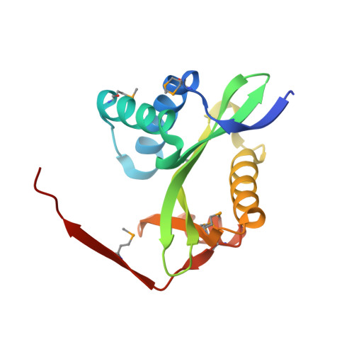 | |
UniProt | |||||
Find proteins for Q185U9 (Clostridioides difficile (strain 630)) Explore Q185U9 Go to UniProtKB: Q185U9 | |||||
Entity Groups | |||||
| Sequence Clusters | 30% Identity50% Identity70% Identity90% Identity95% Identity100% Identity | ||||
| UniProt Group | Q185U9 | ||||
Sequence AnnotationsExpand | |||||
| |||||
| Ligands 2 Unique | |||||
|---|---|---|---|---|---|
| ID | Chains | Name / Formula / InChI Key | 2D Diagram | 3D Interactions | |
| BET Query on BET | D [auth A], F [auth B] | TRIMETHYL GLYCINE C5 H12 N O2 KWIUHFFTVRNATP-UHFFFAOYSA-O |  | ||
| SO4 Query on SO4 | C [auth A], E [auth B] | SULFATE ION O4 S QAOWNCQODCNURD-UHFFFAOYSA-L |  | ||
| Modified Residues 1 Unique | |||||
|---|---|---|---|---|---|
| ID | Chains | Type | Formula | 2D Diagram | Parent |
| MSE Query on MSE | A, B | L-PEPTIDE LINKING | C5 H11 N O2 Se |  | MET |
| Length ( Å ) | Angle ( ˚ ) |
|---|---|
| a = 41.488 | α = 90 |
| b = 66.765 | β = 90 |
| c = 133.884 | γ = 90 |
| Software Name | Purpose |
|---|---|
| REFMAC | refinement |
| SBC-Collect | data collection |
| HKL-3000 | data reduction |
| HKL-3000 | data scaling |
| SHELXD | phasing |
| MLPHARE | phasing |
| DM | phasing |
| RESOLVE | phasing |
| HKL-3000 | phasing |
RCSB PDB (citation) is hosted by
RCSB PDB is a member of the

