3DOP

Crystal structure of 5beta-reductase (AKR1D1) in complex with NADP+ and 5beta-dihydrotestosterone, Resolution 2.00A


Experimental Data Snapshot

  • Method: X-RAY DIFFRACTION
  • Resolution: 2.00 Å
  • R-Value Free: 0.257 
  • R-Value Work: 0.220 
  • R-Value Observed: 0.220 

wwPDB Validation   3D Report Full Report


Ligand Structure Quality Assessment 


This is version 1.2 of the entry. See complete history


Literature

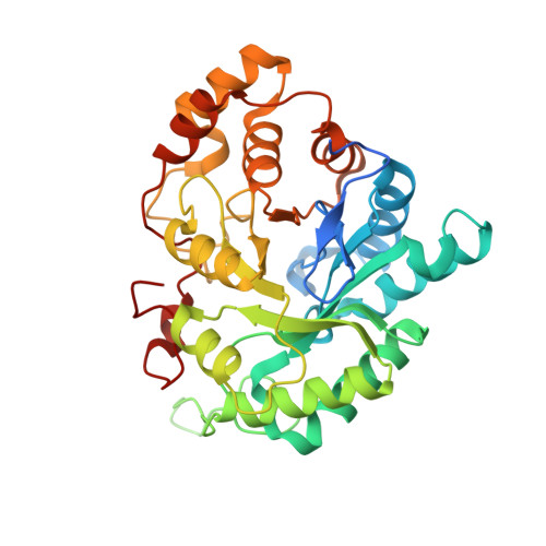
Structure and catalytic mechanism of human steroid 5beta-reductase (AKR1D1)

Di Costanzo, L.Drury, J.E.Christianson, D.W.Penning, T.M.

(2009) Mol Cell Endocrinol 301: 191-198

  • DOI: https://doi.org/10.1016/j.mce.2008.09.013
  • Primary Citation of Related Structures:  
    3DOP

  • PubMed Abstract: 

    Human steroid 5beta-reductase (aldo-keto reductase (AKR) 1D1) catalyzes reduction of Delta(4)-ene double bonds in steroid hormones and bile acid precursors. We have reported the structures of an AKR1D1-NADP(+) binary complex, and AKR1D1-NADP(+)-cortisone, AKR1D1-NADP(+)-progesterone and AKR1D1-NADP(+)-testosterone ternary complexes at high resolutions. Recently, structures of AKR1D1-NADP(+)-5beta-dihydroprogesterone complexes showed that the product is bound unproductively. Two quite different mechanisms of steroid double bond reduction have since been proposed. However, site-directed mutagenesis supports only one mechanism. In this mechanism, the 4-pro-R hydride is transferred from the re-face of the nicotinamide ring to C5 of the steroid substrate. E120, a unique substitution in the AKR catalytic tetrad, permits a deeper penetration of the steroid substrate into the active site to promote optimal reactant positioning. It participates with Y58 to create a "superacidic" oxyanion hole for polarization of the C3 ketone. A role for K87 in the proton relay proposed using the AKR1D1-NADP(+)-5beta-dihydroprogesterone structure is not supported.


  • Organizational Affiliation

    Roy and Diana Vagelos Laboratories, Department of Chemistry, University of Pennsylvania, Philadelphia, PA 19104-6323, USA.


Macromolecules
Find similar proteins by:  (by identity cutoff)  |  3D Structure
Entity ID: 1
MoleculeChains Sequence LengthOrganismDetailsImage
3-oxo-5-beta-steroid 4-dehydrogenase
A, B
346Homo sapiensMutation(s): 0 
Gene Names: AKR1D1SRD5B1
EC: 1.3.1.3
UniProt & NIH Common Fund Data Resources
Find proteins for P51857 (Homo sapiens)
Explore P51857 
Go to UniProtKB:  P51857
PHAROS:  P51857
GTEx:  ENSG00000122787 
Entity Groups  
Sequence Clusters30% Identity50% Identity70% Identity90% Identity95% Identity100% Identity
UniProt GroupP51857
Sequence Annotations
Expand
  • Reference Sequence
Small Molecules
Experimental Data & Validation

Experimental Data

  • Method: X-RAY DIFFRACTION
  • Resolution: 2.00 Å
  • R-Value Free: 0.257 
  • R-Value Work: 0.220 
  • R-Value Observed: 0.220 
  • Space Group: P 21 21 21
Unit Cell:
Length ( Å )Angle ( ˚ )
a = 49.872α = 90
b = 109.495β = 90
c = 129.199γ = 90
Software Package:
Software NamePurpose
CNSrefinement
CrystalCleardata collection
CrystalCleardata reduction
CrystalCleardata scaling
CNSphasing

Structure Validation

View Full Validation Report


Ligand Structure Quality Assessment 


Entry History 

Deposition Data

Revision History  (Full details and data files)

  • Version 1.0: 2008-10-28
    Type: Initial release
  • Version 1.1: 2011-07-13
    Changes: Version format compliance
  • Version 1.2: 2023-11-01
    Changes: Data collection, Database references, Derived calculations, Refinement description