3DML

Crystal structure of the periplasmic thioredoxin SoxS from Paracoccus pantotrophus (reduced form)


Experimental Data Snapshot

  • Method: X-RAY DIFFRACTION
  • Resolution: 1.90 Å
  • R-Value Free: 0.201 
  • R-Value Work: 0.154 
  • R-Value Observed: 0.157 

wwPDB Validation   3D Report Full Report


This is version 1.2 of the entry. See complete history


Literature

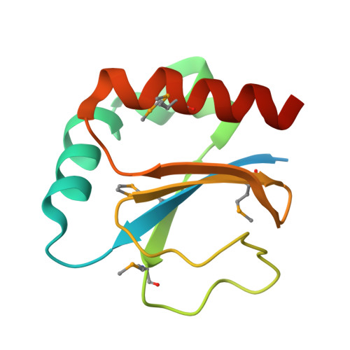
Crystal structure of the periplasmic thioredoxin SoxS from Paracoccus pantotrophus indicates a dual Trx/Grx functionality for activation of chemotrophic sulfur oxidation in vivo

Carius, Y.Rother, D.Friedrich, C.G.Scheidig, A.J.

To be published.

Macromolecules
Find similar proteins by:  (by identity cutoff)  |  3D Structure
Entity ID: 1
MoleculeChains Sequence LengthOrganismDetailsImage
Putative uncharacterized protein116Paracoccus denitrificansMutation(s): 0 
Gene Names: soxS
EC: 1.8.4
UniProt
Find proteins for Q8KM22 (Paracoccus denitrificans)
Explore Q8KM22 
Go to UniProtKB:  Q8KM22
Entity Groups  
Sequence Clusters30% Identity50% Identity70% Identity90% Identity95% Identity100% Identity
UniProt GroupQ8KM22
Sequence Annotations
Expand
  • Reference Sequence
Small Molecules
Modified Residues  1 Unique
IDChains TypeFormula2D DiagramParent
MSE
Query on MSE
A
L-PEPTIDE LINKINGC5 H11 N O2 SeMET
Experimental Data & Validation

Experimental Data

  • Method: X-RAY DIFFRACTION
  • Resolution: 1.90 Å
  • R-Value Free: 0.201 
  • R-Value Work: 0.154 
  • R-Value Observed: 0.157 
  • Space Group: P 31 2 1
Unit Cell:
Length ( Å )Angle ( ˚ )
a = 80.909α = 90
b = 80.909β = 90
c = 33.212γ = 120
Software Package:
Software NamePurpose
REFMACrefinement
MAR345data collection
DENZOdata reduction
SCALEPACKdata scaling
Auto-Rickshawphasing

Structure Validation

View Full Validation Report


Entry History 

Deposition Data

Revision History  (Full details and data files)

  • Version 1.0: 2008-08-26
    Type: Initial release
  • Version 1.1: 2011-07-13
    Changes: Version format compliance
  • Version 1.2: 2017-10-25
    Changes: Refinement description