3DK1

Wild Type HIV-1 Protease with potent Antiviral inhibitor GRL-0105A


Experimental Data Snapshot

  • Method: X-RAY DIFFRACTION
  • Resolution: 1.07 Å
  • R-Value Free: 0.177 
  • R-Value Work: 0.150 
  • R-Value Observed: 0.152 

wwPDB Validation   3D Report Full Report


Ligand Structure Quality Assessment 


This is version 1.5 of the entry. See complete history


Literature

Potent HIV-1 protease inhibitors incorporating meso-bicyclic urethanes as P2-ligands: structure-based design, synthesis, biological evaluation and protein-ligand X-ray studies

Ghosh, A.K.Gemma, S.Takayama, J.Baldridge, A.Leshchenko-Yashchuk, S.Miller, H.B.Wang, Y.F.Kovalevsky, A.Y.Koh, Y.Weber, I.T.Mitsuya, H.

(2008) Org Biomol Chem 6: 3703-3713

  • DOI: https://doi.org/10.1039/b809178a
  • Primary Citation of Related Structures:  
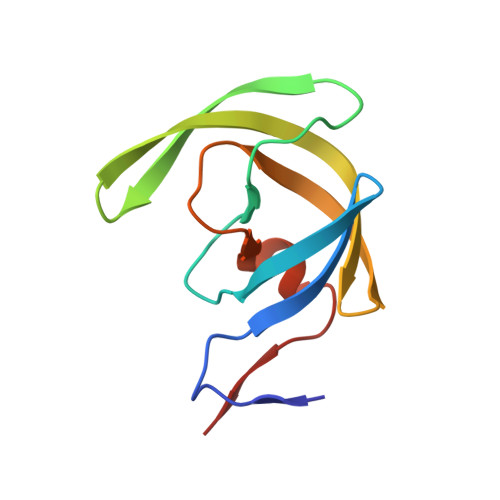
    3DK1

  • PubMed Abstract: 

    Recently, we designed a series of novel HIV-1 protease inhibitors incorporating a stereochemically defined bicyclic fused cyclopentyl (Cp-THF) urethane as the high affinity P2-ligand. Inhibitor with this P2-ligand has shown very impressive potency against multi-drug-resistant clinical isolates. Based upon the -bound HIV-1 protease X-ray structure, we have now designed and synthesized a number of meso-bicyclic ligands which can conceivably interact similarly to the Cp-THF ligand. The design of meso-ligands is quite attractive as they do not contain any stereocenters. Inhibitors incorporating urethanes of bicyclic-1,3-dioxolane and bicyclic-1,4-dioxane have shown potent enzyme inhibitory and antiviral activities. Inhibitor (K(i) = 0.11 nM; IC(50) = 3.8 nM) displayed very potent antiviral activity in this series. While inhibitor showed comparable enzyme inhibitory activity (K(i) = 0.18 nM) its antiviral activity (IC(50) = 170 nM) was significantly weaker than inhibitor . Inhibitor maintained an antiviral potency against a series of multi-drug resistant clinical isolates comparable to amprenavir. A protein-ligand X-ray structure of -bound HIV-1 protease revealed a number of key hydrogen bonding interactions at the S2-subsite. We have created an active model of inhibitor based upon this X-ray structure.


  • Organizational Affiliation

    Departments of Chemistry and Medicinal Chemistry, Purdue University, West Lafayette, IN 47907, USA. akghosh@purdue.edu


Macromolecules
Find similar proteins by:  (by identity cutoff)  |  3D Structure
Entity ID: 1
MoleculeChains Sequence LengthOrganismDetailsImage
Protease
A, B
99Human immunodeficiency virus type 1 (BRU ISOLATE)Mutation(s): 5 
Gene Names: gag-pol
EC: 3.4.23.16
UniProt
Find proteins for P03367 (Human immunodeficiency virus type 1 group M subtype B (isolate BRU/LAI))
Explore P03367 
Go to UniProtKB:  P03367
Entity Groups  
Sequence Clusters30% Identity50% Identity70% Identity90% Identity95% Identity100% Identity
UniProt GroupP03367
Sequence Annotations
Expand
  • Reference Sequence
Small Molecules
Binding Affinity Annotations 
IDSourceBinding Affinity
G05 PDBBind:  3DK1 Ki: 0.18 (nM) from 1 assay(s)
Binding MOAD:  3DK1 Ki: 0.18 (nM) from 1 assay(s)
Experimental Data & Validation

Experimental Data

  • Method: X-RAY DIFFRACTION
  • Resolution: 1.07 Å
  • R-Value Free: 0.177 
  • R-Value Work: 0.150 
  • R-Value Observed: 0.152 
  • Space Group: P 21 21 2
Unit Cell:
Length ( Å )Angle ( ˚ )
a = 58.003α = 90
b = 86.338β = 90
c = 45.833γ = 90
Software Package:
Software NamePurpose
AMoREphasing
SHELXL-97refinement
MAR345data collection
HKL-2000data reduction
HKL-2000data scaling

Structure Validation

View Full Validation Report


Ligand Structure Quality Assessment 


Entry History 

Deposition Data

Revision History  (Full details and data files)

  • Version 1.0: 2009-05-12
    Type: Initial release
  • Version 1.1: 2011-07-13
    Changes: Version format compliance
  • Version 1.2: 2017-08-23
    Changes: Data collection
  • Version 1.3: 2017-10-25
    Changes: Refinement description
  • Version 1.4: 2021-11-10
    Changes: Database references, Derived calculations
  • Version 1.5: 2023-11-01
    Changes: Data collection, Refinement description