3DHW

Crystal structure of methionine importer MetNI


Experimental Data Snapshot

  • Method: X-RAY DIFFRACTION
  • Resolution: 3.70 Å
  • R-Value Free: 0.347 
  • R-Value Work: 0.310 
  • R-Value Observed: 0.310 

wwPDB Validation   3D Report Full Report


This is version 1.2 of the entry. See complete history


Literature

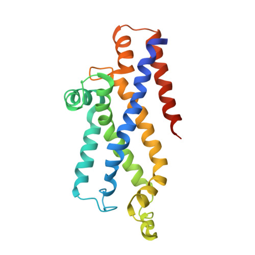
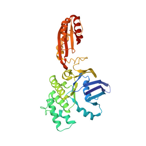
The high-affinity E. coli methionine ABC transporter: structure and allosteric regulation.

Kadaba, N.S.Kaiser, J.T.Johnson, E.Lee, A.Rees, D.C.

(2008) Science 321: 250-253

  • DOI: https://doi.org/10.1126/science.1157987
  • Primary Citation of Related Structures:  
    3DHW, 3DHX

  • PubMed Abstract: 

    The crystal structure of the high-affinity Escherichia coli MetNI methionine uptake transporter, a member of the adenosine triphosphate (ATP)-binding cassette (ABC) family, has been solved to 3.7 angstrom resolution. The overall architecture of MetNI reveals two copies of the adenosine triphosphatase (ATPase) MetN in complex with two copies of the transmembrane domain MetI, with the transporter adopting an inward-facing conformation exhibiting widely separated nucleotide binding domains. Each MetI subunit is organized around a core of five transmembrane helices that correspond to a subset of the helices observed in the larger membrane-spanning subunits of the molybdate (ModBC) and maltose (MalFGK) ABC transporters. In addition to the conserved nucleotide binding domain of the ABC family, MetN contains a carboxyl-terminal extension with a ferredoxin-like fold previously assigned to a conserved family of regulatory ligand-binding domains. These domains separate the nucleotide binding domains and would interfere with their association required for ATP binding and hydrolysis. Methionine binds to the dimerized carboxyl-terminal domain and is shown to inhibit ATPase activity. These observations are consistent with an allosteric regulatory mechanism operating at the level of transport activity, where increased intracellular levels of the transported ligand stabilize an inward-facing, ATPase-inactive state of MetNI to inhibit further ligand translocation into the cell.


  • Organizational Affiliation

    Howard Hughes Medical Institute and Division of Chemistry and Chemical Engineering, Mail Code 114-96, California Institute of Technology, Pasadena, CA 91125, USA.


Macromolecules
Find similar proteins by:  (by identity cutoff)  |  3D Structure
Entity ID: 1
MoleculeChains Sequence LengthOrganismDetailsImage
D-methionine transport system permease protein metI
A, B, E, F
217Escherichia coli K-12Mutation(s): 0 
Gene Names: metIyaeEb0198JW0194
Membrane Entity: Yes 
UniProt
Find proteins for P31547 (Escherichia coli (strain K12))
Explore P31547 
Go to UniProtKB:  P31547
Entity Groups  
Sequence Clusters30% Identity50% Identity70% Identity90% Identity95% Identity100% Identity
UniProt GroupP31547
Sequence Annotations
Expand
  • Reference Sequence
Find similar proteins by:  (by identity cutoff)  |  3D Structure
Entity ID: 2
MoleculeChains Sequence LengthOrganismDetailsImage
Methionine import ATP-binding protein metN
C, D, G, H
343Escherichia coli K-12Mutation(s): 0 
Gene Names: metNabcb0199JW0195
EC: 3.6.3
Membrane Entity: Yes 
UniProt
Find proteins for P30750 (Escherichia coli (strain K12))
Explore P30750 
Go to UniProtKB:  P30750
Entity Groups  
Sequence Clusters30% Identity50% Identity70% Identity90% Identity95% Identity100% Identity
UniProt GroupP30750
Sequence Annotations
Expand
  • Reference Sequence
Experimental Data & Validation

Experimental Data

  • Method: X-RAY DIFFRACTION
  • Resolution: 3.70 Å
  • R-Value Free: 0.347 
  • R-Value Work: 0.310 
  • R-Value Observed: 0.310 
  • Space Group: P 21 21 21
Unit Cell:
Length ( Å )Angle ( ˚ )
a = 97.7α = 90
b = 165.4β = 90
c = 289.02γ = 90
Software Package:
Software NamePurpose
CNSrefinement
Blu-Icedata collection
MOSFLMdata reduction
SCALAdata scaling
SHARPphasing

Structure Validation

View Full Validation Report


Entry History 

Deposition Data

Revision History  (Full details and data files)

  • Version 1.0: 2008-08-05
    Type: Initial release
  • Version 1.1: 2011-07-13
    Changes: Source and taxonomy, Version format compliance
  • Version 1.2: 2024-02-21
    Changes: Data collection, Database references