3DHV

Crystal structure of DltA protein in complex with D-alanine adenylate


Experimental Data Snapshot

  • Method: X-RAY DIFFRACTION
  • Resolution: 2.00 Å
  • R-Value Free: 0.252 
  • R-Value Work: 0.213 
  • R-Value Observed: 0.213 

wwPDB Validation   3D Report Full Report


Ligand Structure Quality Assessment 


This is version 1.3 of the entry. See complete history


Literature

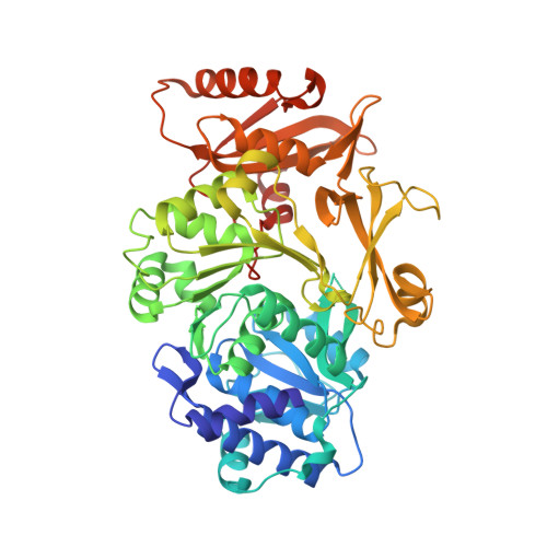
Crystal structure and enantiomer selection by D-alanyl carrier protein ligase DltA from Bacillus cereus.

Du, L.He, Y.Luo, Y.

(2008) Biochemistry 47: 11473-11480

  • DOI: https://doi.org/10.1021/bi801363b
  • Primary Citation of Related Structures:  
    3DHV

  • PubMed Abstract: 

    Ubiquitous D-alanylation of lipoteichoic acids modulates the surface charge and ligand binding of the gram-positive cell wall. Disruption of the bacterial DltABCD gene involved in teichoic acid alanylation, as well as inhibition of the DltA protein, has been shown to increase a gram-positive bacterium's susceptibility to antibiotics. The DltA D-alanyl carrier protein ligase promotes a two-step process starting with adenylation of D-alanine. We have determined the 2.0 A resolution crystal structure of a DltA protein from Bacillus cereus in complex with the D-alanine adenylate intermediate of the first reaction. Despite the low level of sequence similarity, the DltA structure resembles known structures of adenylation domains such as the acetyl-CoA synthetase. The enantiomer selection appears to be enhanced by the medium-sized side chain of Cys-269. The Ala-269 mutant protein shows marked loss of such selection. The network of noncovalent interactions between the D-alanine adenylate and DltA provides structure-based rationale for aiding the design of tight-binding DltA inhibitors for combating infectious gram-positive bacteria such as the notorious methicillin-resistant Staphylococcus aureus.


  • Organizational Affiliation

    Department of Biochemistry, University of Saskatchewan, A3 Health Sciences Building, 107 Wiggins Road, Saskatoon, Saskatchewan, Canada S7N 5E5.


Macromolecules
Find similar proteins by:  (by identity cutoff)  |  3D Structure
Entity ID: 1
MoleculeChains Sequence LengthOrganismDetailsImage
D-alanine-poly(phosphoribitol) ligase512Bacillus cereus ATCC 14579Mutation(s): 1 
Gene Names: dltA
EC: 6.1.1.13
UniProt
Find proteins for Q81G39 (Bacillus cereus (strain ATCC 14579 / DSM 31 / CCUG 7414 / JCM 2152 / NBRC 15305 / NCIMB 9373 / NCTC 2599 / NRRL B-3711))
Explore Q81G39 
Go to UniProtKB:  Q81G39
Entity Groups  
Sequence Clusters30% Identity50% Identity70% Identity90% Identity95% Identity100% Identity
UniProt GroupQ81G39
Sequence Annotations
Expand
  • Reference Sequence
Small Molecules
Ligands 2 Unique
IDChains Name / Formula / InChI Key2D Diagram3D Interactions
AMP
Query on AMP

Download Ideal Coordinates CCD File 
C [auth A]ADENOSINE MONOPHOSPHATE
C10 H14 N5 O7 P
UDMBCSSLTHHNCD-KQYNXXCUSA-N
DAL
Query on DAL

Download Ideal Coordinates CCD File 
B [auth A]D-ALANINE
C3 H7 N O2
QNAYBMKLOCPYGJ-UWTATZPHSA-N
Binding Affinity Annotations 
IDSourceBinding Affinity
DAL PDBBind:  3DHV Kd: 1.10e+6 (nM) from 1 assay(s)
Experimental Data & Validation

Experimental Data

  • Method: X-RAY DIFFRACTION
  • Resolution: 2.00 Å
  • R-Value Free: 0.252 
  • R-Value Work: 0.213 
  • R-Value Observed: 0.213 
  • Space Group: P 1 21 1
Unit Cell:
Length ( Å )Angle ( ˚ )
a = 60.7α = 90
b = 86.7β = 113.5
c = 57.4γ = 90
Software Package:
Software NamePurpose
PROTEUM PLUSdata collection
AMoREphasing
CNSrefinement
PROTEUM PLUSdata reduction
PROTEUM PLUSdata scaling

Structure Validation

View Full Validation Report


Ligand Structure Quality Assessment 


Entry History 

Deposition Data

Revision History  (Full details and data files)

  • Version 1.0: 2008-11-04
    Type: Initial release
  • Version 1.1: 2011-07-13
    Changes: Version format compliance
  • Version 1.2: 2021-10-20
    Changes: Database references, Derived calculations
  • Version 1.3: 2023-08-30
    Changes: Data collection, Refinement description