3DGV

Crystal structure of thrombin activatable fibrinolysis inhibitor (TAFI)


Experimental Data Snapshot

  • Method: X-RAY DIFFRACTION
  • Resolution: 2.50 Å
  • R-Value Free: 0.243 
  • R-Value Work: 0.205 

wwPDB Validation   3D Report Full Report


Ligand Structure Quality Assessment 


This is version 2.2 of the entry. See complete history


Literature

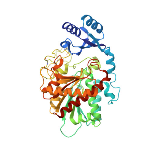
The crystal structure of thrombin-activable fibrinolysis inhibitor (TAFI) provides the structural basis for its intrinsic activity and the short half-life of TAFIa.

Anand, K.Pallares, I.Valnickova, Z.Christensen, T.Vendrell, J.Wendt, K.U.Schreuder, H.A.Enghild, J.J.Aviles, F.X.

(2008) J Biol Chem 283: 29416-29423

  • DOI: https://doi.org/10.1074/jbc.M804003200
  • Primary Citation of Related Structures:  
    3DGV

  • PubMed Abstract: 

    Mature thrombin-activable fibrinolysis inhibitor (TAFIa) is a highly unstable metallocarboxypeptidase that stabilizes blood clots by clipping C-terminal lysine residues from partially degraded fibrin. In accordance with its in vitro antifibrinolytic activity, animal studies have reported that inhibition of mature TAFI aids in the prevention of thrombosis. The level of TAFI activity is stringently regulated through (i) controlled proteolytic truncation of the zymogen (TAFI), generating the mature enzyme, TAFIa, and (ii) the short half-life of TAFIa. TAFI itself exhibits an intrinsic enzymatic activity, which is likely required to provide a baseline level of antifibrinolytic activity. The novel crystal structure presented here reveals that the active site of TAFI is accessible, providing the structural explanation for the its intrinsic activity. It also supports the notion that an "instability region" exists, in agreement with site-directed mutagenesis studies. Sulfate ions, bound to this region, point toward a potential heparin-binding site and could explain how heparin stabilizes TAFIa.


  • Organizational Affiliation

    Sanofi-Aventis Pharma Deutschland GmbH, Industriepark Höchst, 65926 Frankfurt am Main, Germany.


Macromolecules
Find similar proteins by:  (by identity cutoff)  |  3D Structure
Entity ID: 1
MoleculeChains Sequence LengthOrganismDetailsImage
Carboxypeptidase B2
A, B, C
401Bos taurusMutation(s): 0 
EC: 3.4.17.20
UniProt
Find proteins for Q2KIG3 (Bos taurus)
Explore Q2KIG3 
Go to UniProtKB:  Q2KIG3
Entity Groups  
Sequence Clusters30% Identity50% Identity70% Identity90% Identity95% Identity100% Identity
UniProt GroupQ2KIG3
Sequence Annotations
Expand
  • Reference Sequence
Oligosaccharides

Help

Entity ID: 2
MoleculeChains Length2D Diagram Glycosylation3D Interactions
2-acetamido-2-deoxy-alpha-D-glucopyranose-(1-4)-2-acetamido-2-deoxy-beta-D-glucopyranose
D, I
2N-Glycosylation
Glycosylation Resources
GlyTouCan:  G07375KG
GlyCosmos:  G07375KG
GlyGen:  G07375KG
Entity ID: 3
MoleculeChains Length2D Diagram Glycosylation3D Interactions
beta-L-fucopyranose-(1-6)-2-acetamido-2-deoxy-beta-D-glucopyranose
E
2N-Glycosylation
Glycosylation Resources
GlyTouCan:  G61843VN
GlyCosmos:  G61843VN
GlyGen:  G61843VN
Entity ID: 4
MoleculeChains Length2D Diagram Glycosylation3D Interactions
2-acetamido-2-deoxy-beta-D-glucopyranose-(1-4)-2-acetamido-2-deoxy-beta-D-glucopyranose
F, J
2N-Glycosylation
Glycosylation Resources
GlyTouCan:  G42666HT
GlyCosmos:  G42666HT
GlyGen:  G42666HT
Entity ID: 5
MoleculeChains Length2D Diagram Glycosylation3D Interactions
2-acetamido-2-deoxy-beta-D-glucopyranose-(1-4)-[beta-L-fucopyranose-(1-6)]2-acetamido-2-deoxy-beta-D-glucopyranose
G
3N-Glycosylation
Glycosylation Resources
GlyTouCan:  G28454KX
GlyCosmos:  G28454KX
GlyGen:  G28454KX
Entity ID: 6
MoleculeChains Length2D Diagram Glycosylation3D Interactions
beta-D-mannopyranose-(1-4)-2-acetamido-2-deoxy-beta-D-glucopyranose-(1-4)-2-acetamido-2-deoxy-beta-D-glucopyranose
H
3N-Glycosylation
Glycosylation Resources
GlyTouCan:  G15407YE
GlyCosmos:  G15407YE
GlyGen:  G15407YE
Small Molecules
Ligands 4 Unique
IDChains Name / Formula / InChI Key2D Diagram3D Interactions
NAG
Query on NAG

Download Ideal Coordinates CCD File 
JA [auth C],
V [auth B]
2-acetamido-2-deoxy-beta-D-glucopyranose
C8 H15 N O6
OVRNDRQMDRJTHS-FMDGEEDCSA-N
MLI
Query on MLI

Download Ideal Coordinates CCD File 
IA [auth B],
NA [auth C],
U [auth A]
MALONATE ION
C3 H2 O4
OFOBLEOULBTSOW-UHFFFAOYSA-L
SO4
Query on SO4

Download Ideal Coordinates CCD File 
AA [auth B]
BA [auth B]
CA [auth B]
DA [auth B]
EA [auth B]
AA [auth B],
BA [auth B],
CA [auth B],
DA [auth B],
EA [auth B],
FA [auth B],
GA [auth B],
HA [auth B],
L [auth A],
LA [auth C],
M [auth A],
MA [auth C],
N [auth A],
O [auth A],
P [auth A],
Q [auth A],
R [auth A],
S [auth A],
T [auth A],
X [auth B],
Y [auth B],
Z [auth B]
SULFATE ION
O4 S
QAOWNCQODCNURD-UHFFFAOYSA-L
ZN
Query on ZN

Download Ideal Coordinates CCD File 
K [auth A],
KA [auth C],
W [auth B]
ZINC ION
Zn
PTFCDOFLOPIGGS-UHFFFAOYSA-N
Experimental Data & Validation

Experimental Data

  • Method: X-RAY DIFFRACTION
  • Resolution: 2.50 Å
  • R-Value Free: 0.243 
  • R-Value Work: 0.205 
  • Space Group: P 41 21 2
Unit Cell:
Length ( Å )Angle ( ˚ )
a = 146.47α = 90
b = 146.47β = 90
c = 231.69γ = 90
Software Package:
Software NamePurpose
DNAdata collection
PHASERphasing
CNSrefinement
XDSdata reduction
XSCALEdata scaling

Structure Validation

View Full Validation Report


Ligand Structure Quality Assessment 


Entry History 

Deposition Data

Revision History  (Full details and data files)

  • Version 1.0: 2008-07-22
    Type: Initial release
  • Version 1.1: 2011-07-13
    Changes: Non-polymer description, Version format compliance
  • Version 1.2: 2017-02-08
    Changes: Other
  • Version 2.0: 2020-07-29
    Type: Remediation
    Reason: Carbohydrate remediation
    Changes: Advisory, Atomic model, Data collection, Derived calculations, Structure summary
  • Version 2.1: 2021-04-28
    Changes: Database references, Structure summary
  • Version 2.2: 2023-11-01
    Changes: Data collection, Database references, Refinement description