3DGO

A non-biological ATP binding protein with a Tyr-Phe mutation in the ligand binding domain


Experimental Data Snapshot

  • Method: X-RAY DIFFRACTION
  • Resolution: 2.50 Å
  • R-Value Free: 0.230 
  • R-Value Work: 0.184 
  • R-Value Observed: 0.186 

wwPDB Validation   3D Report Full Report


Ligand Structure Quality Assessment 


This is version 1.3 of the entry. See complete history


Literature

A synthetic protein selected for ligand binding affinity mediates ATP hydrolysis.

Simmons, C.R.Stomel, J.M.McConnell, M.D.Smith, D.A.Watkins, J.L.Allen, J.P.Chaput, J.C.

(2009) ACS Chem Biol 4: 649-658

  • DOI: https://doi.org/10.1021/cb900109w
  • Primary Citation of Related Structures:  
    3DGL, 3DGN, 3DGO

  • PubMed Abstract: 

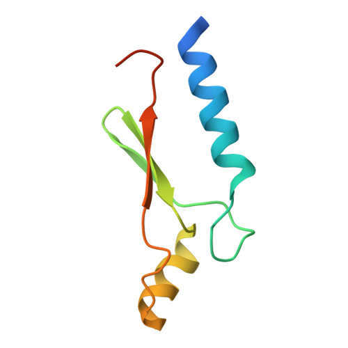
    How primitive enzymes emerged from a primordial pool remains a fundamental unanswered question with important practical implications in synthetic biology. Here we show that a de novo evolved ATP binding protein, selected solely on the basis of its ability to bind ATP, mediates the regiospecific hydrolysis of ATP to ADP when crystallized with 1 equiv of ATP. Structural insights into this reaction were obtained by growing protein crystals under saturating ATP conditions. The resulting crystal structure refined to 1.8 A resolution reveals that this man-made protein binds ATP in an unusual bent conformation that is metal-independent and held in place by a key bridging water molecule. Removal of this interaction using a null mutant results in a variant that binds ATP in a normal linear geometry and is incapable of ATP hydrolysis. Biochemical analysis, including high-resolution mass spectrometry performed on dissolved protein crystals, confirms that the reaction is accelerated in the crystalline environment. This observation suggests that proteins with weak chemical reactivity can emerge from high affinity ligand binding sites and that constrained ligand-binding geometries could have helped to facilitate the emergence of early protein enzymes.


  • Organizational Affiliation

    Center for BioOptical Nanotechnology, The Biodesign Institute, Tempe, Arizona 85287-5201, USA.


Macromolecules
Find similar proteins by:  (by identity cutoff)  |  3D Structure
Entity ID: 1
MoleculeChains Sequence LengthOrganismDetailsImage
ATP Binding Protein-DX81unidentifiedMutation(s): 0 
Entity Groups  
Sequence Clusters30% Identity50% Identity70% Identity90% Identity95% Identity100% Identity
Sequence Annotations
Expand
  • Reference Sequence
Small Molecules
Binding Affinity Annotations 
IDSourceBinding Affinity
ATP Binding MOAD:  3DGO Kd: 180 (nM) from 1 assay(s)
PDBBind:  3DGO Kd: 180 (nM) from 1 assay(s)
Experimental Data & Validation

Experimental Data

  • Method: X-RAY DIFFRACTION
  • Resolution: 2.50 Å
  • R-Value Free: 0.230 
  • R-Value Work: 0.184 
  • R-Value Observed: 0.186 
  • Space Group: P 32 2 1
Unit Cell:
Length ( Å )Angle ( ˚ )
a = 71.742α = 90
b = 71.742β = 90
c = 55.488γ = 120
Software Package:
Software NamePurpose
DENZOdata reduction
SCALEPACKdata scaling
PHASERphasing
DMphasing
REFMACrefinement
PDB_EXTRACTdata extraction
HKL-2000data scaling

Structure Validation

View Full Validation Report


Ligand Structure Quality Assessment 


Entry History 

Deposition Data

Revision History  (Full details and data files)

  • Version 1.0: 2009-06-30
    Type: Initial release
  • Version 1.1: 2011-07-13
    Changes: Version format compliance
  • Version 1.2: 2017-10-25
    Changes: Refinement description
  • Version 1.3: 2024-02-21
    Changes: Data collection, Database references, Derived calculations